Lots of interesting abstracts and cases were submitted for TCTAP 2023. Below are the accepted ones after a thorough review by our official reviewers. Don’t miss the opportunity to expand your knowledge and interact with authors as well as virtual participants by sharing your opinion in the comment section!
TCTAP C-188
Transcatheter Closure of Ruptured Sinus of Valsalva With PDA Occluder Device in Previously Surgically Repaired Ventricular Septal Defect
By Atul Kaushik, Surender Deora
Presenter
Atul Kaushik
Authors
Atul Kaushik1, Surender Deora1
Affiliation
All India Institute of Medical Sciences, Jodhpur, India1,
View Study Report
TCTAP C-188
STRUCTURAL HEART DISEASE - Others (Structural Heart Disease)
Transcatheter Closure of Ruptured Sinus of Valsalva With PDA Occluder Device in Previously Surgically Repaired Ventricular Septal Defect
Atul Kaushik1, Surender Deora1
All India Institute of Medical Sciences, Jodhpur, India1,
Clinical Information
Patient initials or Identifier Number
Mr X
Relevant Clinical History and Physical Exam
18 year old boy operated surgically 3 years back for Sub aortic Ventricular septal defect at other center.Now presented with complaints of Dyspnea on effort NYHA III x 4 months.On Examination Blood Pressure: 120/50 mm HgHear rate: 78/minJVP : Not raised, No Pedal edema/Clubbing/cyanosis.Surgical Scar of median sternotomy present.CVS: S1 , S2 : NormalA continuous murmur heard over Left parasternal area in 2nd and 3rd intercostal space.No LVS3/S4, No pericardial rub.
Relevant Test Results Prior to Catheterization
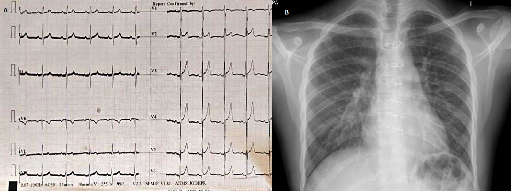
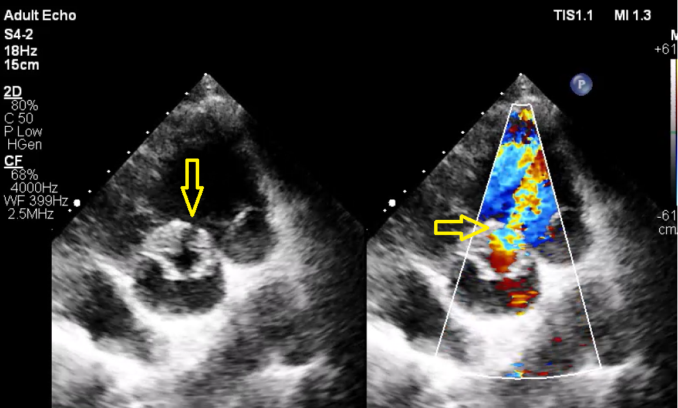
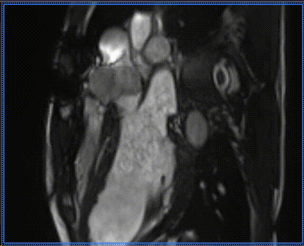
ECG: Normal sinus rhythm with Left ventricular hypertrophy. (Picture 1)Chest X ray: Normal CT ratio with increased pulmonary blood flow. (Picture 1)TTE: Mildly dilated left ventricle, No shunt across Ventricular septum. A 6 mm defect noted from Right coronary sinus to right ventricle without any aneurysm (yellow arrow). No AR/ MR/TR. Normal LV function, Ejection fraction ~60 %. (Picture 2)Cardiac MRI: 6.2 mm Defect noted from Right coronary sinus to RV (white arrow, picture 3).


Relevant Catheterization Findings
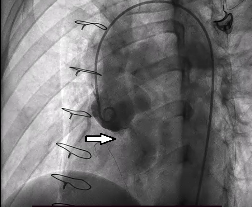
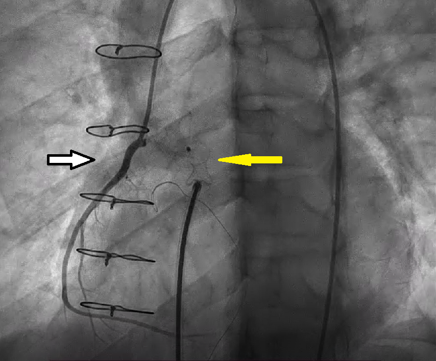
Aortogram was done through right femoral approach with 6 French pigtail catheter in LAO Cranial and RAO views (Picture 4) which showed ruptured sinus of valsalva with flow towards right ventricular outflow tract a defect of size 6.5 mm (white arrow) with Right coronary artery away from the site of rupture.

Interventional Management
Procedural Step
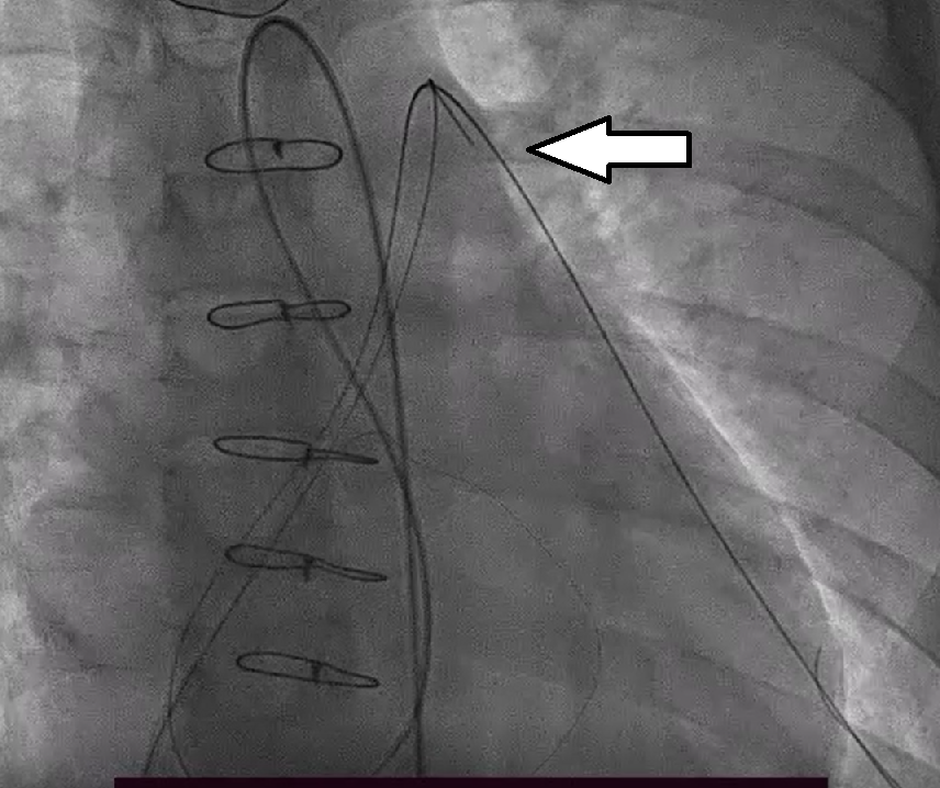
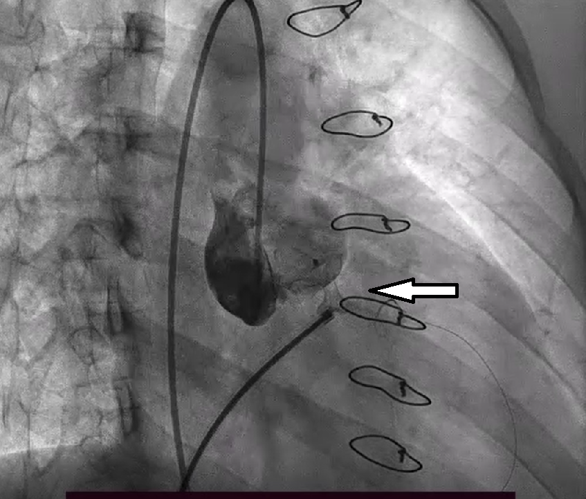
Patient was planned for device closure with the use of PDA occluder. Femoral vein access was taken with 7 Fr and arterial access was taken with 6 French sheaths. A J tipped hydrophilic wire was used and the defect was crossed from aorta to RVOT with the help of judkins right 5 Fr catheter. The wire was placed in the left pulmonary artery (LPA) and then through the right femoral vein access the wire was snared from the LPA with the help of a snare, and the access to defect was gained from the venous side. (Picture 5). A 8/10 PDA occluder device (Lifetech) was deployed after confirming that the device is away from the origin of right coronary artery (Picture 6). After performing the aortogram and confirming the position the device was deployed(Picture 7).


Case Summary
Sinus of valsalva rupture is mostly congenital, however it may also be acquired. In our case, we believe that it was acquired as there was no defect at the time of previous surgery. On echocardiography there was lack of aneurysm formation/ "wind sock" appearance, however cardiac MRI may corroborate and may prove important while planning these cases. Device therapies with the use of PDA/VSD occluder device may be done without complications when planned properly.


