Lots of interesting abstracts and cases were submitted for TCTAP 2023. Below are the accepted ones after a thorough review by our official reviewers. Don’t miss the opportunity to expand your knowledge and interact with authors as well as virtual participants by sharing your opinion in the comment section!
TCTAP C-017
A Case Report: A Case of TGCV With Restenosis in a Short Period of Time.
By Kento Kikuchi, Yusuke Nakano, Atomu Tajima, Masanobu Fujimoto, Akihiro Suzuki, Hirohiko Ando, Hiroaki Takashima, Tetsuya Amano
Presenter
Kento Kikuchi
Authors
Kento Kikuchi1, Yusuke Nakano1, Atomu Tajima1, Masanobu Fujimoto1, Akihiro Suzuki1, Hirohiko Ando1, Hiroaki Takashima1, Tetsuya Amano1
Affiliation
Aichi Medical University, Japan1,
View Study Report
TCTAP C-017
CORONARY - Acute Coronary Syndromes (STEMI, NSTE-ACS)
A Case Report: A Case of TGCV With Restenosis in a Short Period of Time.
Kento Kikuchi1, Yusuke Nakano1, Atomu Tajima1, Masanobu Fujimoto1, Akihiro Suzuki1, Hirohiko Ando1, Hiroaki Takashima1, Tetsuya Amano1
Aichi Medical University, Japan1,
Clinical Information
Patient initials or Identifier Number
K.O
Relevant Clinical History and Physical Exam
A 76-y-o female visited our hospital becauseof chest pain lasting for 30 minutes. Angina was suspected and admitted to thehospital for close examination. He had a history of PCI for ACS with a DESfor the treatment ofLAD proximal lesion 4 months ago, including type 2 diabetes and dyslipidemia.She doesn't have a smoking history. The therapeuticagents were administered: aspirin, and statins. Physical examination: Blood pressure 134/70mmhg; pulse 59bpm; No cardiac murmur, no rale no leg edema.
Relevant Test Results Prior to Catheterization
No apparent abnormality, exceptfor HbA1c 6.0 % and creatinine 0.76 mg/dL, were detected by blood tests. ChestX-ray showed no effusions in the lung fields and the normal rangecardiothoracic ratio. ECG showed QS pattern in V1-3 and ST depression in V4-6.Echocardiography shows hypokinesis in the anterior wall. Eventually, she wassuspected of cardiac ischemia and received CAG.
Relevant Catheterization Findings
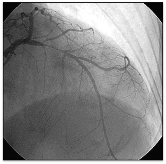
Diffuse in-stent restenosis was observed in patients with TGCV.
Interventional Management
Procedural Step
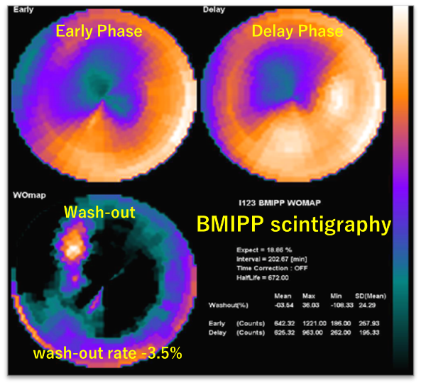
Coronary angiography revealeddiffuse in-stent restenosis (ISR) in LAD (#6 90%) with diffuse mild narrowingcoronary vessels. Balloon dilation was performed to ISR and TIMI 3 coronaryflow were obtained. The present case presented with diffuse type ISR, even withDES. These findings suggest a rather different mechanism of ISR other than theconventional risk factors of ISR, and we performed 123I-β-methyl-P-iodophenyl-pentadecanoicacids (BMIPP) scintigraphy, which detect abnormal fatty acid metabolism, forclose examination of risk factors of ISR. BMIPP scintigraphy showed aremarkably decreased washout rate of less than 10 %. Based on diffuse mildnarrowing coronary vessels and decreased wash-out rate of less than 10 % inBMIPP scintigraph, we diagnosed this case as triglyceride depositcardiomyovasculopathy (TGCV). TGCV is a novel disease concept characterized byan excessive accumulation of triglyceride in cardiomyocytes and vascular smoothmuscle cells, leading to coronary artery disease. TGCV has been reported to be an increased risk for vascular failure after DES implantation due to excessiveinflammatory cytokines and growth factors, leading to a diffuse occlusiverestenosis morphology, even after the implantation of DES.


Case Summary
Here, we present a case of TGCVwith specific restenosis morphology following DES implantation. Comorbidity ofTGCV has been reported to be associated with an increased incidence of vascularfailure. Therefore, we should pay attention to TGCV as a risk stratificationaround PCI.


