Lots of interesting abstracts and cases were submitted for TCTAP 2023. Below are the accepted ones after a thorough review by our official reviewers. Don’t miss the opportunity to expand your knowledge and interact with authors as well as virtual participants by sharing your opinion in the comment section!
TCTAP C-139
A Unique Case of in Stents Restenosis: The Rings of Fire
By Yuen Hoong Phang, Vijayendran Rajalingam, Kai Soon Liew, Saravanan Krishinan, Dharmaraj Karthikesan
Presenter
Yuen Hoong Phang
Authors
Yuen Hoong Phang1, Vijayendran Rajalingam2, Kai Soon Liew1, Saravanan Krishinan3, Dharmaraj Karthikesan4
Affiliation
Sultanah Bahiyah Hospital, Malaysia1, Sultan Idris Shah Serdang Hospital, Malaysia2, Ministry of Health Malaysia, Malaysia3, Hospital Sultanah Bahiyah, Malaysia4,
View Study Report
TCTAP C-139
CORONARY - Stents (Bare-metal, Drug-eluting)
A Unique Case of in Stents Restenosis: The Rings of Fire
Yuen Hoong Phang1, Vijayendran Rajalingam2, Kai Soon Liew1, Saravanan Krishinan3, Dharmaraj Karthikesan4
Sultanah Bahiyah Hospital, Malaysia1, Sultan Idris Shah Serdang Hospital, Malaysia2, Ministry of Health Malaysia, Malaysia3, Hospital Sultanah Bahiyah, Malaysia4,
Clinical Information
Patient initials or Identifier Number
AS00952233
Relevant Clinical History and Physical Exam
75 years old Indian gentleman with underlying type 2 diabetes mellitus, hypertension, dyslipidemia, and ischemic heart disease. He had a history of angioplasty done in 2020 and 2021, presented with typical angina, and was treated for acute coronary syndrome. Both procedures were done in different state hospitals, and procedural notes were not available during the intervention. Physical examination was unremarkable. BP 173/80, HR 58, dextrostix 14.2.
Relevant Test Results Prior to Catheterization
ECG showed sinus rhythm with fragmented QRS complexes over inferior leads.Hemoglobin 14.3 / white cell count 6.5 / Platelet 292Urea 4.1 / Sodium 136 / Potassium 4.1 / Creatinine 85Total Cholesterol 4.2 / Triglyceride 1.6 / HDL 1.0 / LDL 2.51ALT 16 / Total Protein 69 / Bilirubin 6
Echocardiogram showed left ventricle hypertrophy, dilated left atrium, no regional wall abnormalities, ejection fraction of 64% with diastolic grade 1.
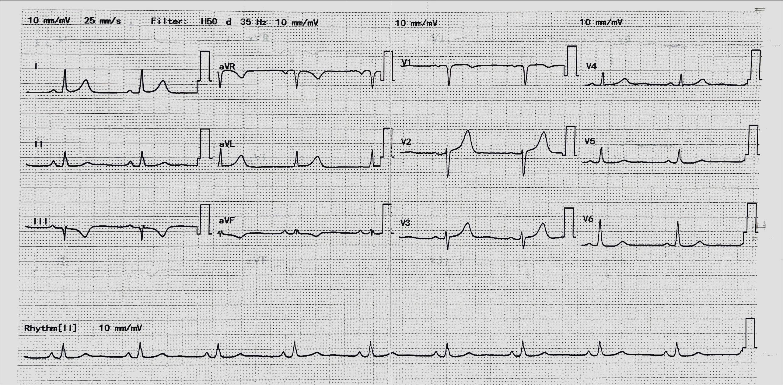
Echocardiogram showed left ventricle hypertrophy, dilated left atrium, no regional wall abnormalities, ejection fraction of 64% with diastolic grade 1.

Relevant Catheterization Findings
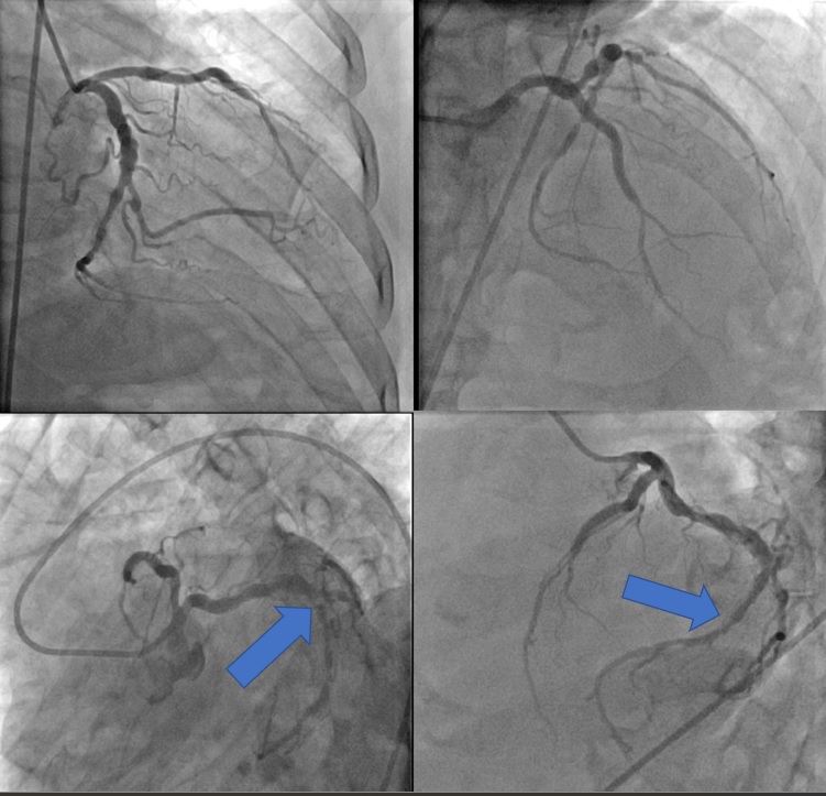
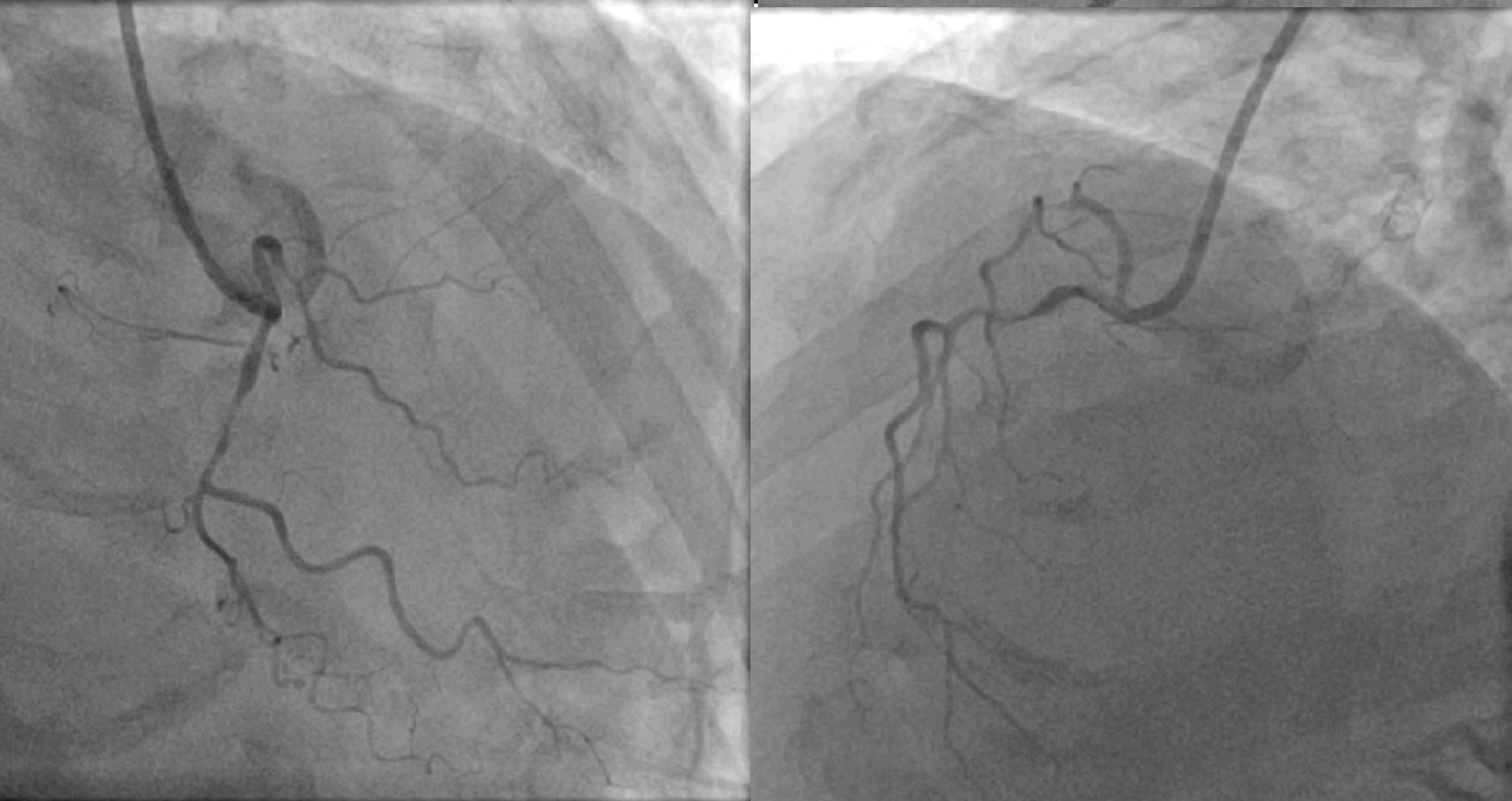
Left dominant system with recessive RCALeft Main is smoothLAD – Proximal LAD stent was patent with mild in-stent stenosis. There was a diffuse disease distal to stent, with 50% stenosis from mid to distal LADLCX – Large in calibre with ectatic vessels. Tight 90-95% stenosis over mid-LCX, best appreciated in LAO Cranial view, with suspicious overlying calcium or stent over the lesion.


Interventional Management
Procedural Step
Access over left femoral as failed right radial and femoral artery cannulation.
Diagnostic angiogram with 6F JL4.0/JR 4.0. Decided to treat LCX.Guider 7F EBU 3.5 to LCA.
Sion Blue wired to LCX and Runthrough Floppy to OM2.
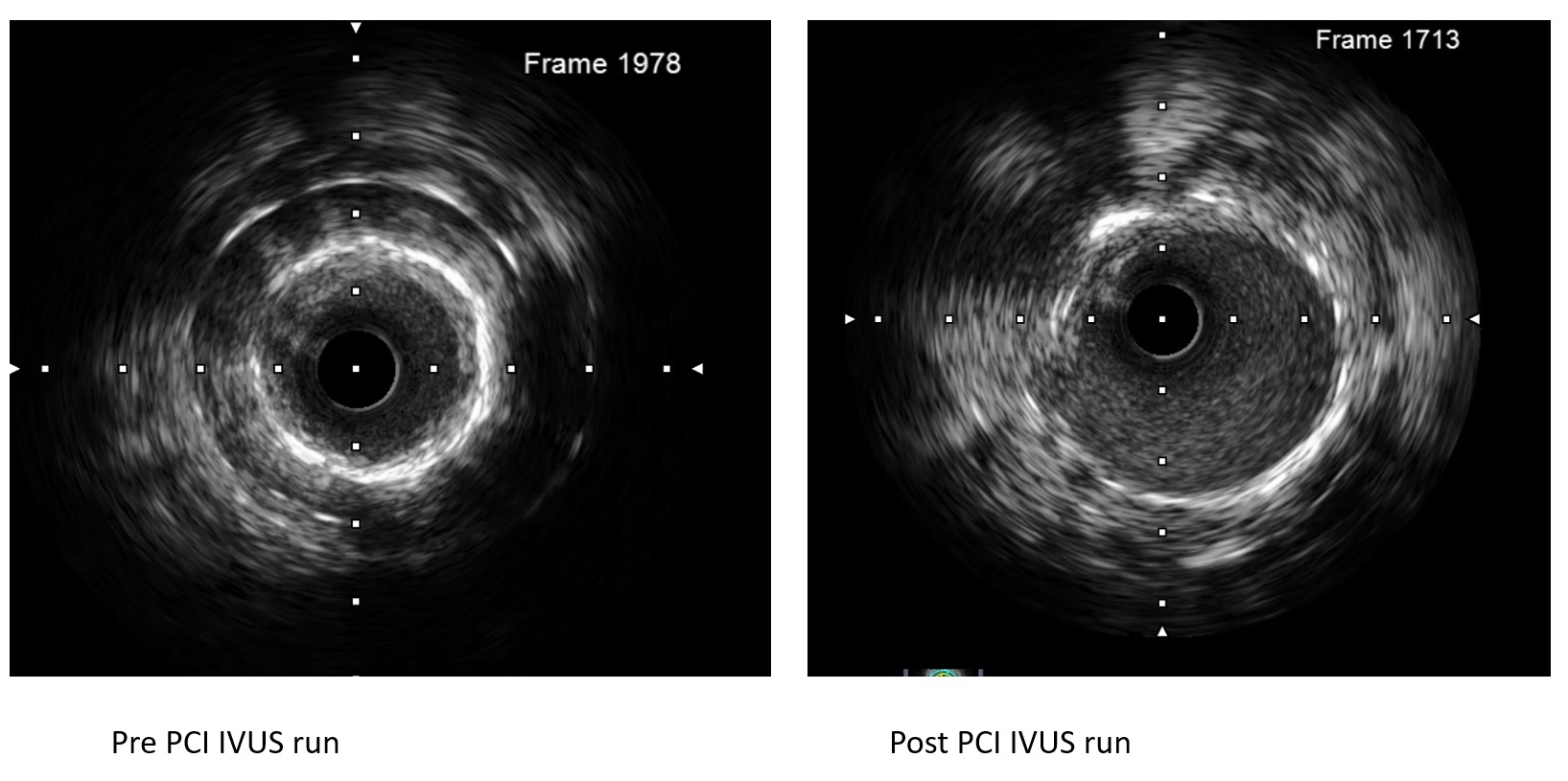
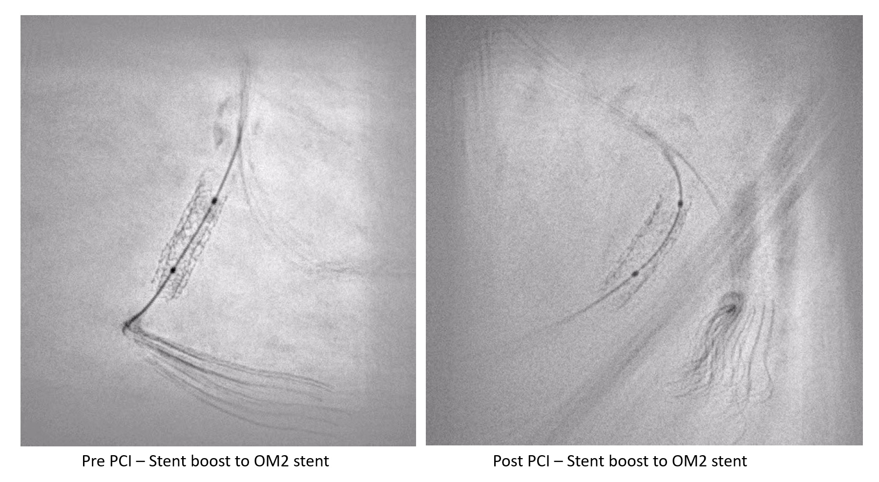
SC Balloon 2.0x15mm was passed down to LCX and a stent boost was done over the suspicious lesion. Noted there were TWO overlapping stents over the same site, with one markedly under-expanded. The lesion was predilated with SC Balloon @ 6atm, 2.00mmIVUS run noted vessel size was about 4.0mm with the first stent well opposed, but the second layer stent was under-expanded with plaque in between the first and second stents and neointimal hyperplasia over the second stent leading to small lumen size with MLA of 3.33mm2 and lumen size of 2.00mm. There was a segment of 360o calcium about 3mm in length. The plaque burden was about 75%.The lesion was sequentially predilated with Scoring Balloon 3.5x15mm @ 12-30atm, ID 3.5-3.92mm. Stent boost noted the second stent was well opposed. IVUS run confirmed it, and noted non-flow limiting dissection over the distal stent edge.
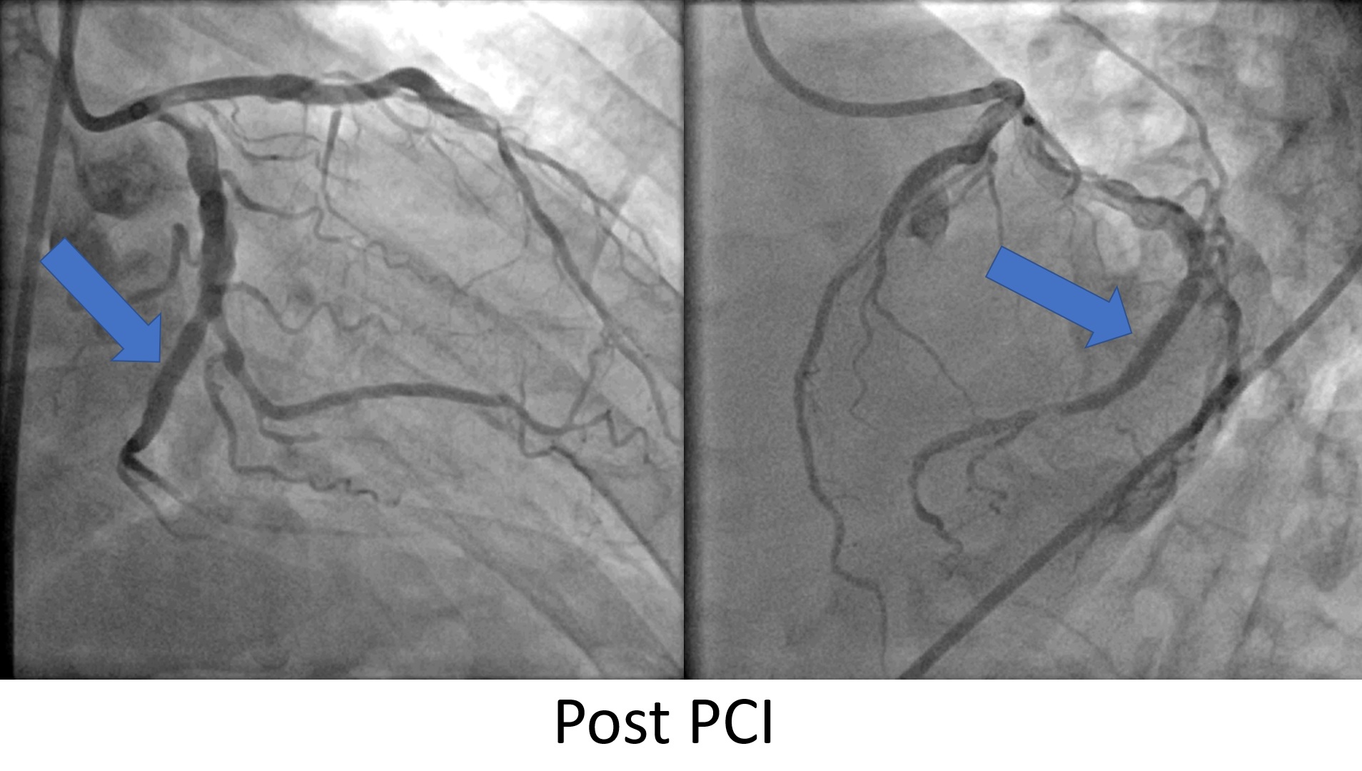
The lesion was further optimized with NC Balloon 3.72@ 15mm @ 24-30atm, ID 3.93 -3.97mm.IVUS run noted good stent opposition and distal stent edge dissection remained similar. Distal stent edge MLA was 7.02mm2 with a vessel size of 4.0mm and proximal lesion MLA was 9.17mm2 with a vessel size of 5.00mm. The lesion length was 27mm. The lesion was treated with DEB Prevail 4.00x30mm @ 11atm, ID 4.14atm for 90 seconds. Post DCB noted good angiographic results with marked luminal gain.He was started on dual antiplatelet, GDMT, and was scheduled for a surveillance angiogram in 6 months.


Case Summary
We postulated that the second PCI operator did not realize that there was a stent in the LCX, as we had difficulty identifying the two-layered stents on a conventional angiogram. Hence, obtaining the previous procedural findings before an intervention is crucial.The etiology of in-stent restenosis for the second stent was likely due to inadequate lesion preparation and under-expansion. The first Sirolimus stent was well opposed with good expansion, hence we suspected the early in-stent restenosis of the first stent was secondary to neointimal hyperplasia and we opted for a Paclitaxel-based DEB.Management of in-stent restenosis is well eluded by Giustino in Coronary Instent Restenosis JACC review.However, the presented case was an uncommon form of in-stent restenosis with under-expansion of the second stent. We were concerned about the risk of coronary perforation, but the lesion was prepared under imaging guidance. The utilization of stent boost and intravascular imaging were vital in this PCI.


