Lots of interesting abstracts and cases were submitted for TCTAP 2023. Below are the accepted ones after a thorough review by our official reviewers. Don’t miss the opportunity to expand your knowledge and interact with authors as well as virtual participants by sharing your opinion in the comment section!
TCTAP C-083
Balloon Won’t Cross, Rotational Atherectomy to Do Away With Coronary Calcification.
By Anindya Banerjee, Shashikant Singh, Subhas Pramanik, Ramachandra Barik
Presenter
Anindya Banerjee
Authors
Anindya Banerjee1, Shashikant Singh1, Subhas Pramanik1, Ramachandra Barik1
Affiliation
All India Institute of Medical Sciences, Bhubaneswar, India1,
View Study Report
TCTAP C-083
CORONARY - Chronic Total Occlusion
Balloon Won’t Cross, Rotational Atherectomy to Do Away With Coronary Calcification.
Anindya Banerjee1, Shashikant Singh1, Subhas Pramanik1, Ramachandra Barik1
All India Institute of Medical Sciences, Bhubaneswar, India1,
Clinical Information
Patient initials or Identifier Number
DB
Relevant Clinical History and Physical Exam
A 37-year-old gentleman with no comorbidities presented with progressively worsening effort angina and dyspnea on exertion since past 4 month. He had a history of anterior wall ST elevation myocardial infarction (STEMI) with delayed presentation 4 months back and was on medical therapy for the same. At presentation physical examination including cardiovascular examination was unremarkable.
Relevant Test Results Prior to Catheterization
An electrocardiogram revealed qRBBB pattern. Echocardiogram showed anteroseptal hypokinesia with a reduced ejection fraction of 44%.Cardiac biomarker - Troponin I kit test was negative at presentation. Rest of the laboratory parameters were within normal limit.
Relevant Catheterization Findings
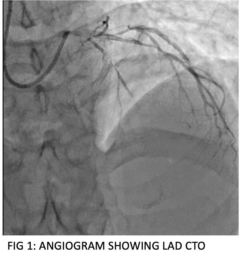
A coronary angiogram revealed bifurcating normal left main coronary artery, chronic total occlusion (CTO) of left anterior descending (LAD) after first diagonal (D1), nondominant left circumflex (LCX) diffuse distal disease and two obtuse marginals (OM) having 50-60% lesion, and dominant right coronary artery (RCA) having CTO of posterior left ventricular branch. Based on these findings after a heart team and informed patient discussion LAD intervention was planned.
Interventional Management
Procedural Step
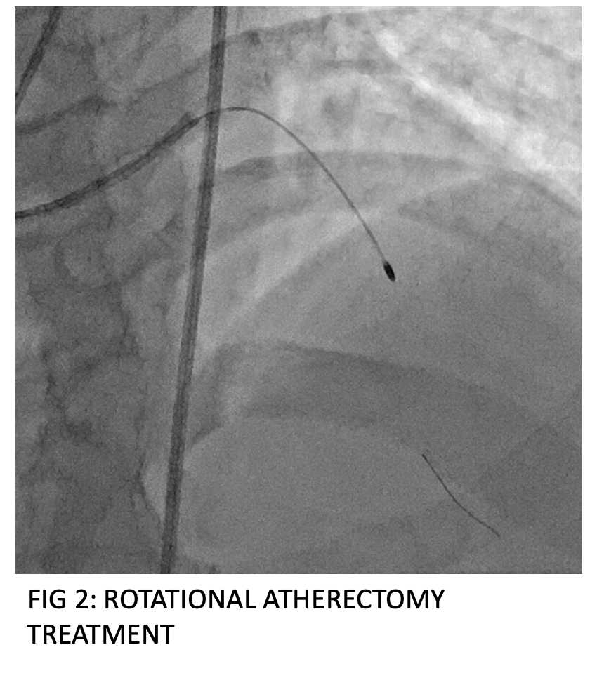
The patient was taken to cardiac catheterization laboratory and through a 6 French (Fr) right radial access the left ostium was engaged with a 6 Fr XB guide. The LAD lesion was crossed with a Fielder XT A guidewire, however, even a 0.85 * 10 mm nic nano balloon could not be crossed across the lesion. The same was tried with a different Fielder FC wire but to no avail. The procedure was abandoned at that point and a reattempt was planned with high speed rotational atherectomy (HSRA).


Case Summary
Angioplasty in a balloon uncrossable lesion poses a unique challenge to the operator. A pragmatic approach to dealing with such lesions at first include increasing guide catheter support (7 Fr or 8 Fr), use of additional guidewire support, using microcatheters, or using low profile (0.85 – 1.5 mm) SC balloon with lubricious coating. Failing these strategies more advanced techniques like atherectomy – laser, HSRA, or orbital can be employed, as demonstrated in this case.


