Lots of interesting abstracts and cases were submitted for TCTAP 2023. Below are the accepted ones after a thorough review by our official reviewers. Don’t miss the opportunity to expand your knowledge and interact with authors as well as virtual participants by sharing your opinion in the comment section!
TCTAP C-181
Pulmonary Valve Balloon Dilation and Transcatheter Closure of Atrial Septal Defect in a Teenaged Cyanotic Girl
By Abdul Momen, Ashraf Ur Rahman, Lima Sayami, ABM Abm Nurunnobi, Md Saqif Shahriar, Md Zulfikar Ali
Presenter
Ashraf Ur Rahman
Authors
Abdul Momen1, Ashraf Ur Rahman1, Lima Sayami2, ABM Abm Nurunnobi1, Md Saqif Shahriar1, Md Zulfikar Ali1
Affiliation
National Institute of Cardiovascular Diseases (NICVD), Bangladesh1, Nicvd, Bangladesh2,
View Study Report
TCTAP C-181
STRUCTURAL HEART DISEASE - Congenital Heart Disease (ASD, PDA, PFO, VSD)
Pulmonary Valve Balloon Dilation and Transcatheter Closure of Atrial Septal Defect in a Teenaged Cyanotic Girl
Abdul Momen1, Ashraf Ur Rahman1, Lima Sayami2, ABM Abm Nurunnobi1, Md Saqif Shahriar1, Md Zulfikar Ali1
National Institute of Cardiovascular Diseases (NICVD), Bangladesh1, Nicvd, Bangladesh2,
Clinical Information
Patient initials or Identifier Number
Miss X
Relevant Clinical History and Physical Exam
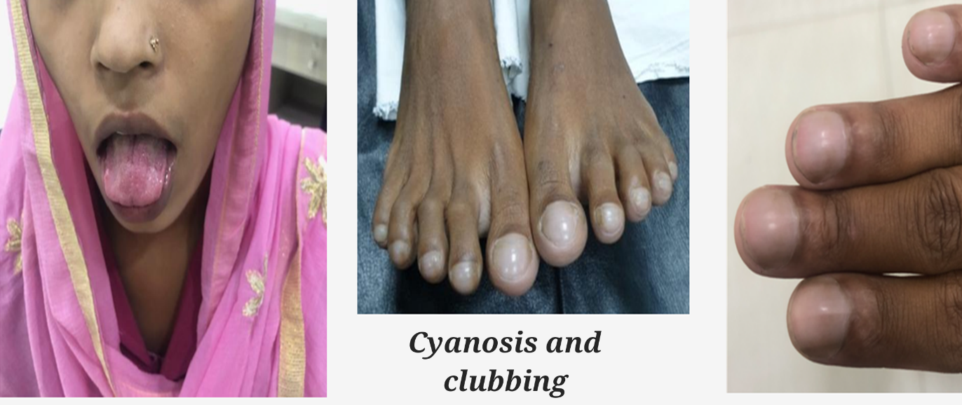
A 18 years short stature married teenaged girl presented with increasing dyspnoea (NYHA II) and palpitation on exertion for last 6 months. She had history of stillbirth 8 months back. Central Cyanosis, digital clubbing and congested conjunctiva were evident. Her cardiac examination revealed splitted 2nd heart sound and a soft 4/6 ejection systolic murmer at upper left sternal border.

Relevant Test Results Prior to Catheterization
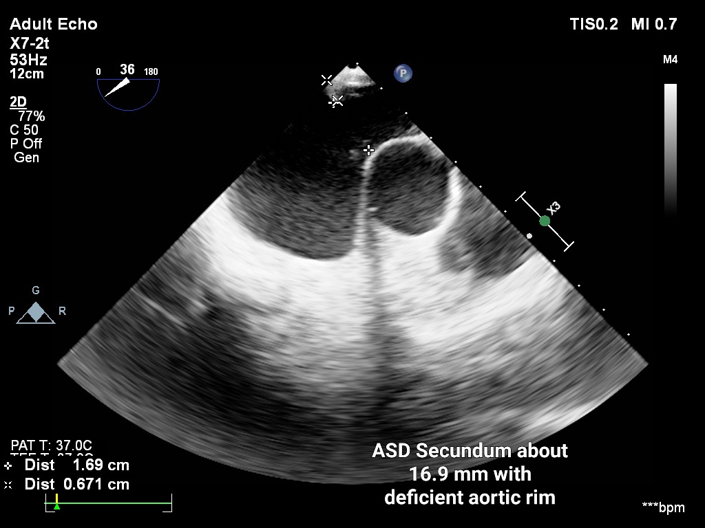
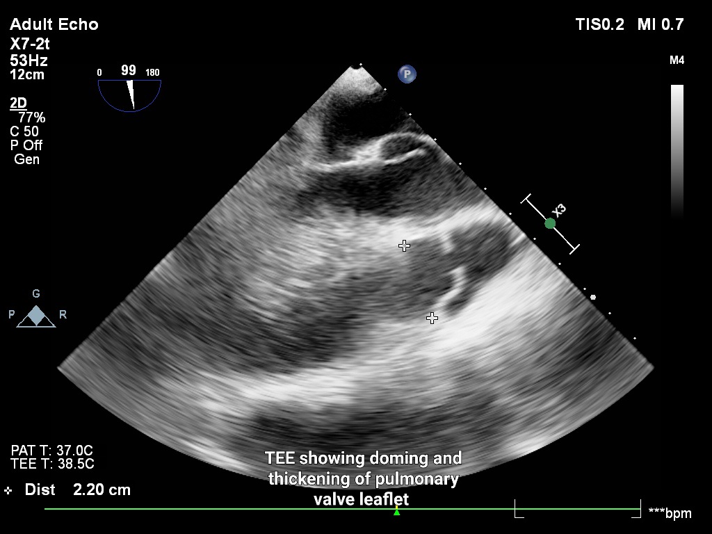
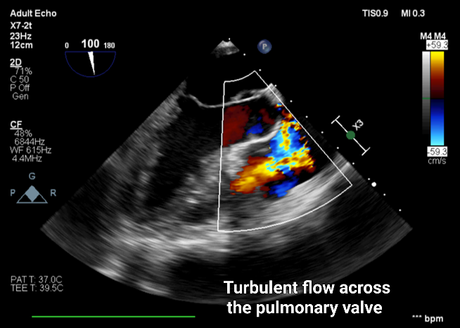
ECG showed incomplete RBBB and RVH with strain. Her SpO2 was 85% at room temperature and Hb was 18 mg/dl. Transthoracic followed by transoesophageal echocardiography revealed large ASD secundum (16x19x17 mm) with bidirectional shunt. Rims were adequate except the aortic rim. Severe pulmonary valvular stenosis with peak systolic pressure gradient of 68 mmHg was found. Pulmonary valve was thickened, mildly calcified and pulmonary annulus was 22 mm. LV and RV function were normal (TAPSE 23mm).


Relevant Catheterization Findings
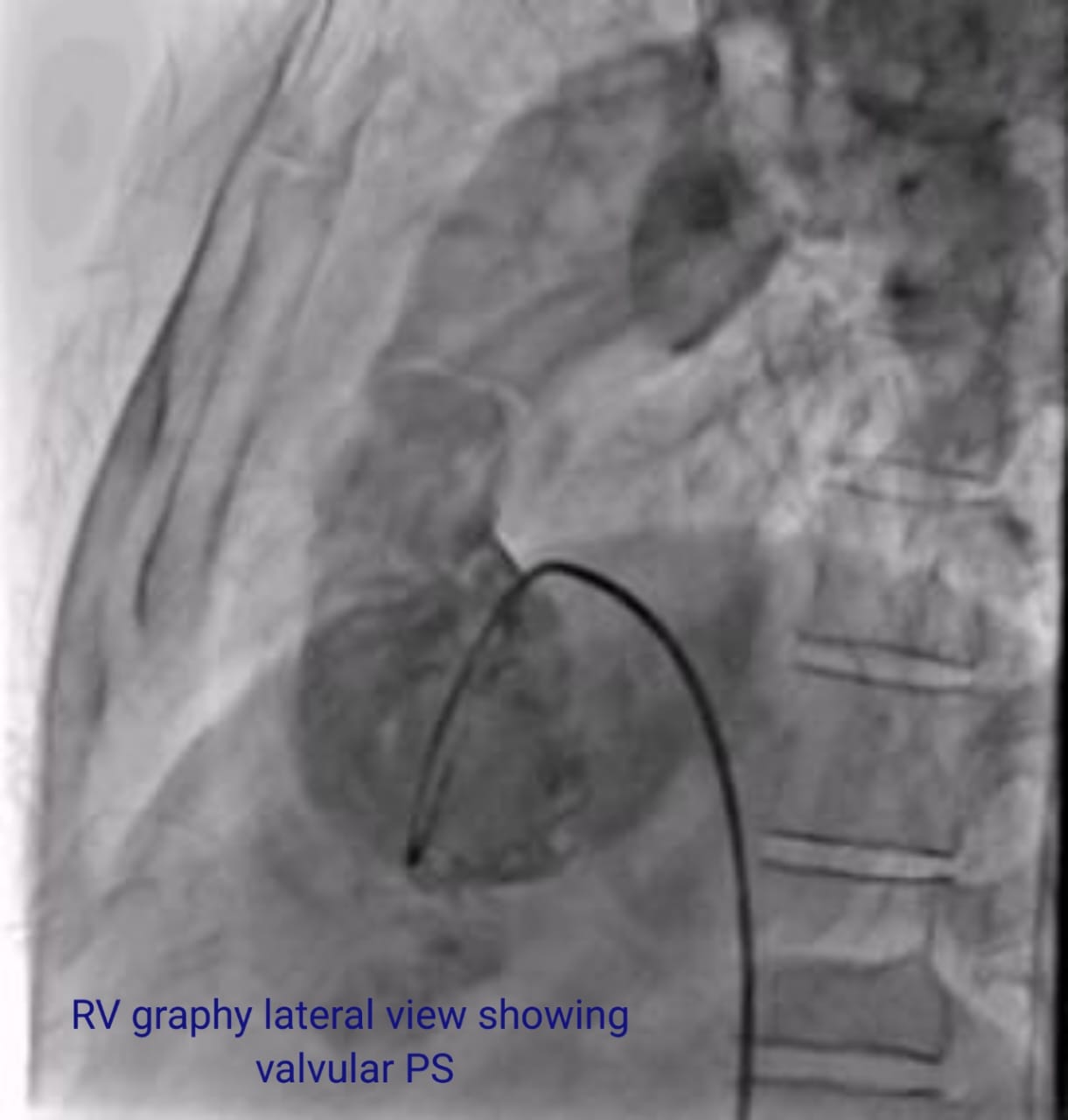
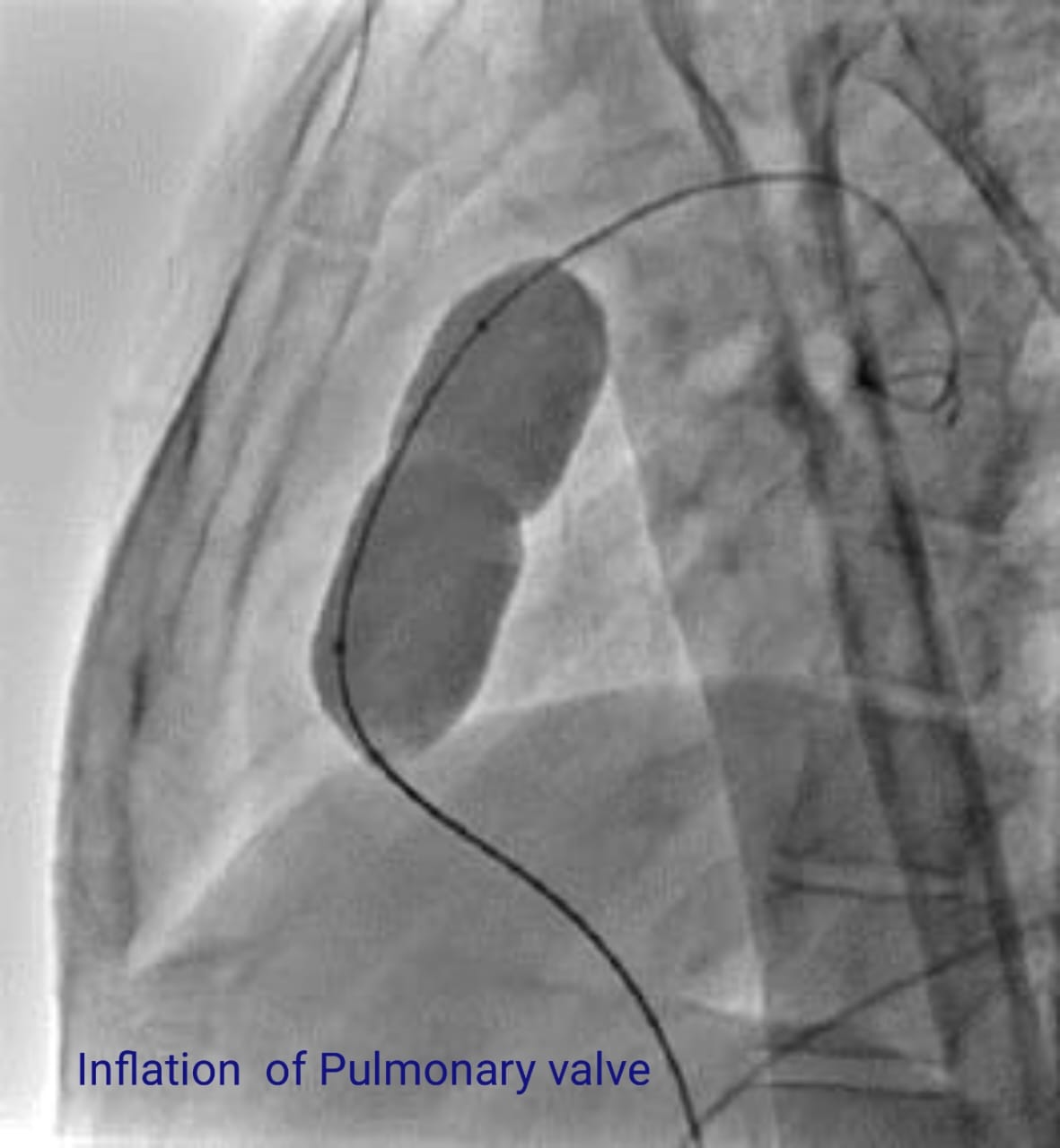
we planned to do pulmonary Valvuloplasty first. RV graphy antero-posterior and lateral views showed severe valvular PS and post stenotic dilation of pulmonary artery. RV peak systolic pressure was 61 mm Hg and PA systolic pressure was 7 mm Hg. So the gradient across pulmonary valve was 54 mm Hg. Pulmonary valve was dilated by 24X50 mm Vulver Balloon and the gradient decreased to 20 mmHg from 54 mmHg. P02 was 88%, Hb dropped from 18 gm/dl to 17 gm/dl. But cyanosis and clubbing were still evident.


Interventional Management
Procedural Step
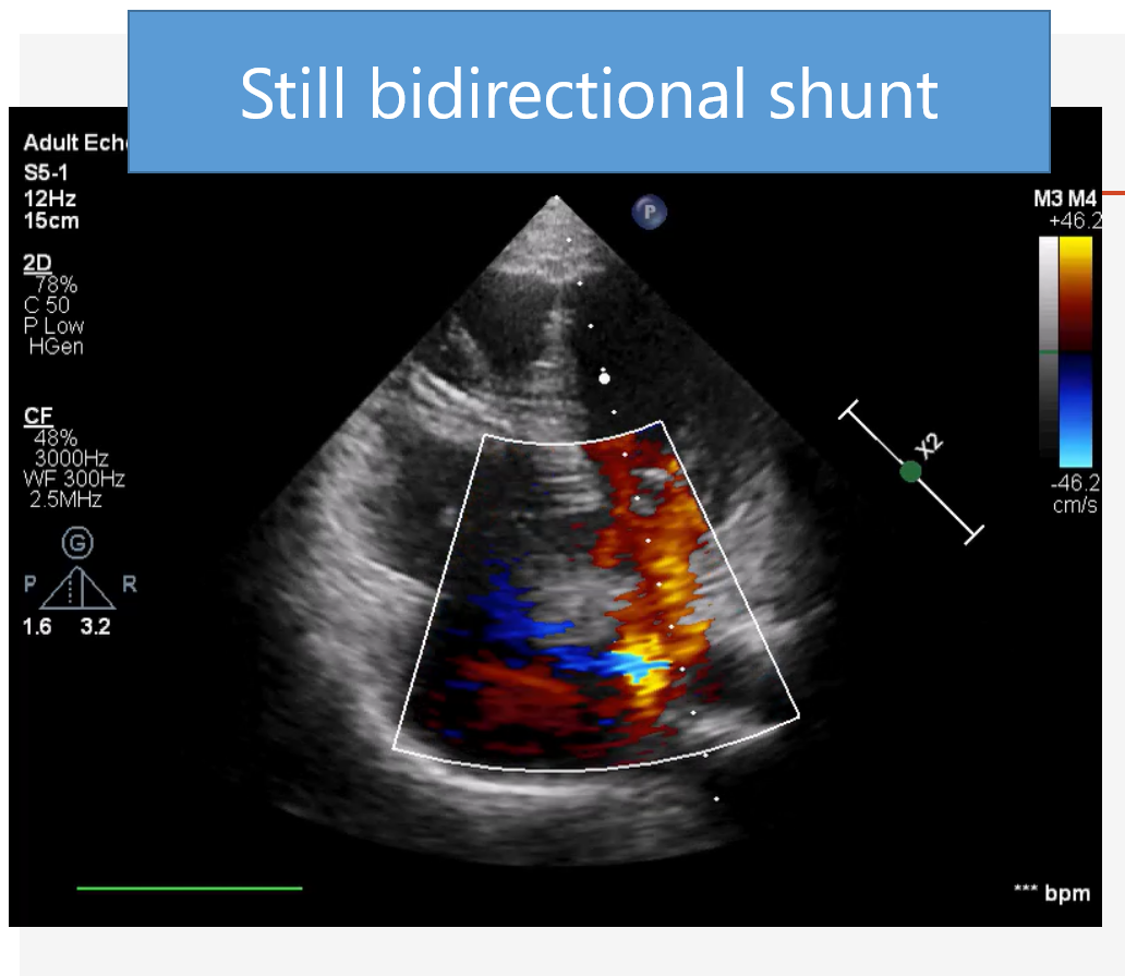
Following pulmonary valvuloplasty the patient symptomatically improved. We waited for about one and half months. Echocardiography revealed no significant gradient across pulmonary valve (28 mm Hg), but shunt was bidirectional across the ASD even though normal PASP (32mmHg). So we decided to do cardiac catheterization and ASD device closure if cath data were favourable.
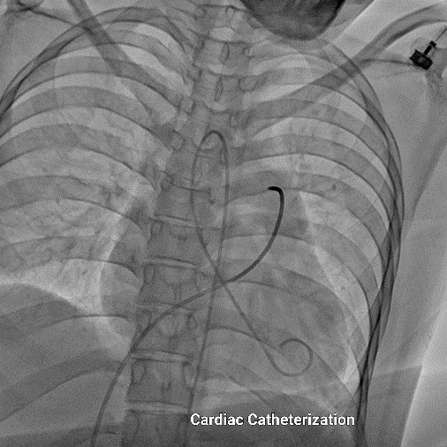
Diagnostic catheterization revealed 6 mmHg of right atrial pressure, 20 mmHg of systolic pulmonary arterial pressure and 4 mmHg pulmonary capillary wedge pressure. Oxygen saturation at lower SVC was 63% & in mid right atrium 76% indicating significant step up. Oxygen saturation at left upper pulmonary vein was 97% & in aorta was 88% indicating significant step down. so there was a bidirectional shunt. the Qp/Qs was calculated as 1.76 and PVR was 1.59 woods unit.
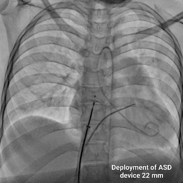
22 mm Amplatzer Septal Occluder was deployed properly with TEE guidance. We did not remove the delivery cable and waited for about 20 minutes. Then we repeated the cardiac cath . After the occlusion of the ASD by device, the aortic saturation was significantly increased reaching to 97%. The right atrial pressure increased to 7 mmHg which was 6 mmHg previously. As the patient was haemodynamically stable and there was no significant (>2 mmHg) rise of RA pressure, we finally removed the delivery cable. The position of the device was reconfirmed by TEE guidance.


Case Summary
Right to left inter atrial shunt is not always a contra indication for correction of the shunt. Elucidation of the actual cause of this shunt is important. The rare combination of ASD with severe PS having bidirectional shunt in adult can be safely corrected by transcatheter approach. The transcatheter approach is an effective and an attractive method in alleviating cyanosis in these patients. The careful selection of the patient and an earlier intervention with resolution of systemic hypoxia lead to the favorable hemodynamic outcome. The stepwise approach is suggested for the treatment of suitable patients


