Lots of interesting abstracts and cases were submitted for TCTAP 2023. Below are the accepted ones after a thorough review by our official reviewers. Don’t miss the opportunity to expand your knowledge and interact with authors as well as virtual participants by sharing your opinion in the comment section!
TCTAP C-192
An Unusual Case of Culotte Type Ruptured Sinus of Valsalva With Two Windsocks Originating From an Aneurysmal Sac
By Shishir Soni, Bhanu Duggal, Anish Gupta
Presenter
Shishir Soni
Authors
Shishir Soni1, Bhanu Duggal2, Anish Gupta2
Affiliation
SSH, NSCB Medical College, India1, AIIMS Rishikesh, India2,
View Study Report
TCTAP C-192
STRUCTURAL HEART DISEASE - Others (Structural Heart Disease)
An Unusual Case of Culotte Type Ruptured Sinus of Valsalva With Two Windsocks Originating From an Aneurysmal Sac
Shishir Soni1, Bhanu Duggal2, Anish Gupta2
SSH, NSCB Medical College, India1, AIIMS Rishikesh, India2,
Clinical Information
Patient initials or Identifier Number
20210066484
Relevant Clinical History and Physical Exam
A 44-year female was referred for the evaluation of sudden onset chest pain preceding the dyspnea and bilateral lower limb oedema for 2 weeks. On physical examination, she had wide pulse pressure and a continuous murmur predominant over the left 4th-5th intercostal space near the sternal margin. A ruptured sinus of Valsalva into the right atrium (RA) was suspected based on the above findings along with screening echocardiography done at the emergency,therefore planned for TEE after stabilisation.

Relevant Test Results Prior to Catheterization
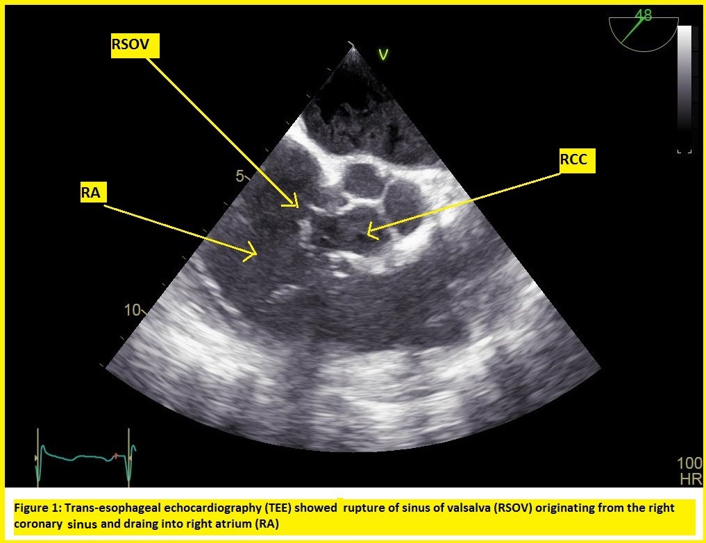
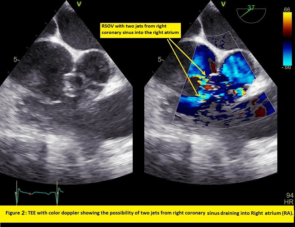
Transesophageal echocardiography (TEE) was performed which showed the Ruptured sinus of Valsalva (RSOV) draining into the RA (Figure 1).Adding colour doppler and on changing the angulation of TEE probe, the possibility of two windsocks originating from the right coronary sinus was raised (Figure 2), although it was unusual and not depicted well in the literature. Therefore, agitated saline contrast was injected into the left arm to delineate the two jets of RSOV on TEE.

Relevant Catheterization Findings
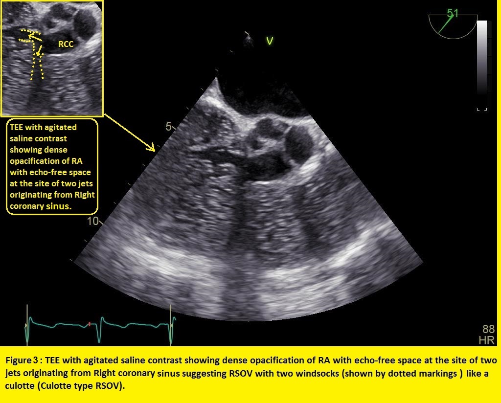
TEE with agitated saline contrast showed dense opacification of RA with echo-free space at the site of two jets originating from the Right coronary sinus suggesting an RSOV with two windsocks like a culotte i.e. Culotte type RSOV (figure 3) which has never been described well in the literature. As this anatomy was not well suited for device closure therefore she was referred for surgical closure. Patient underwent coronary angiography which showed normal epicardial coronaries.

Interventional Management
Procedural Step
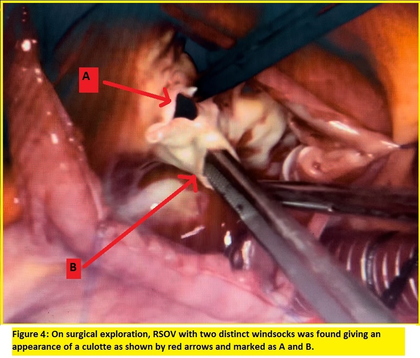
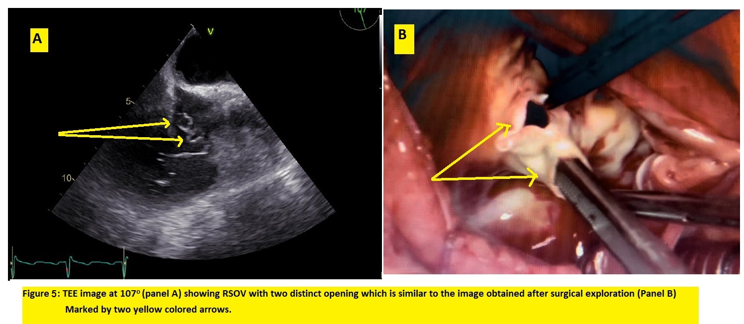
TEE images depicted the unusual Culotte-type RSOV with two distinct openings and therefore it might have been challenging by the percutaneous device closure approach. Although the patient did not have associated Ventricular septal defect or aortic regurgitation, however, due to this rare entity with anatomically distinct type of RSOV, surgical closure was preferred. After a discussion with the institutional heart team, the patient was taken up for the surgical closure of RSOV by the CTVS surgeon. After midline sternotomy, aortic and bi-caval cannulation; cardiopulmonary bypass was instituted. Aortotomy was done and the right atrium opened following which two windsocks were identified arising from the right coronary sinus like a culotte (figure 4) which was suggested in the prior evaluation by TEE with agitated saline contrast. RSOV was successfully repaired with a PTFE patch using a bicameral approach. The Right atrium and aorta were closed. Surprisingly the enface view of the RSOV with two distinct openings evidenced in the surgical field was similar to the TEE images at 1070 angulation (Figure 5) obtained on prior evaluation. Subsequently, she was weaned from the cardiopulmonary bypass. The post-operative period was uneventful and the patient was discharged subsequently.


Case Summary
This is a rare entity and an RSOV with two windsocks that we called a "Culotte type" RSOV has been (almost) never described in the literature.Use of TEE with agitated saline contrast for delineating RSOV morphology although unique but useful in this case. Identifying the exact anatomy before proceeding with the surgical or device closure is the key aspect in the management of such patients. This type of RSOV i.e. culotte type may be added as another subtype, as Sakakibara and Konno’s classification or its modified classification does not take into account this morphology.


