
Lots of interesting abstracts and cases were submitted for TCTAP 2021 Virtual. Below are accepted ones after thoroughly reviewed by our official reviewers. Don’t miss the opportunity to explore your knowledge and interact with authors as well as virtual participants by sharing your opinion!
TCTAP C-099
Presenter
Nailya Mukhamadeeva
Authors
Nailya Mukhamadeeva1, Diana Gareeva2, Valentin Pavlov2, Igor V. Buzaev2, Dmitriy Krekotin1, Dmitry V. Onegov3, Vladimir Plechev2, Irina Evgenievna Nikolaeva3, Shihua Zhao4, Naufal Zagidullin2
Affiliation
International Medical Centre VS Buzaev Memorial, Russian Federation1, Bashkir State Medical University, Russian Federation2, Republican Cardiology Center, Russian Federation3, Cardiovascular Institute and Fuwai Hospital, China4,
View Study Report
TCTAP C-099
IMAGING AND PHYSIOLOGIC LESION ASSESSMENT - Imaging: Non-Invasive
Clinical Case of Asymptomatic Cardiac Subepicardial Interventricular Lipoma
Nailya Mukhamadeeva1, Diana Gareeva2, Valentin Pavlov2, Igor V. Buzaev2, Dmitriy Krekotin1, Dmitry V. Onegov3, Vladimir Plechev2, Irina Evgenievna Nikolaeva3, Shihua Zhao4, Naufal Zagidullin2
International Medical Centre VS Buzaev Memorial, Russian Federation1, Bashkir State Medical University, Russian Federation2, Republican Cardiology Center, Russian Federation3, Cardiovascular Institute and Fuwai Hospital, China4,
Clinical Information
Patient initials or Identifier Number
13YOB
Relevant Clinical History and Physical Exam
A 13-year-old boy was diagnosed with a right ventricular (RV) tumor when he was 5 years old and has no cardiac-related complains and symptoms till the present moment. He was growing according to his age, is studying at the school, and doing sport (biathlon). The hemodynamics was unchanged and stable.
Relevant Test Results Prior to Catheterization
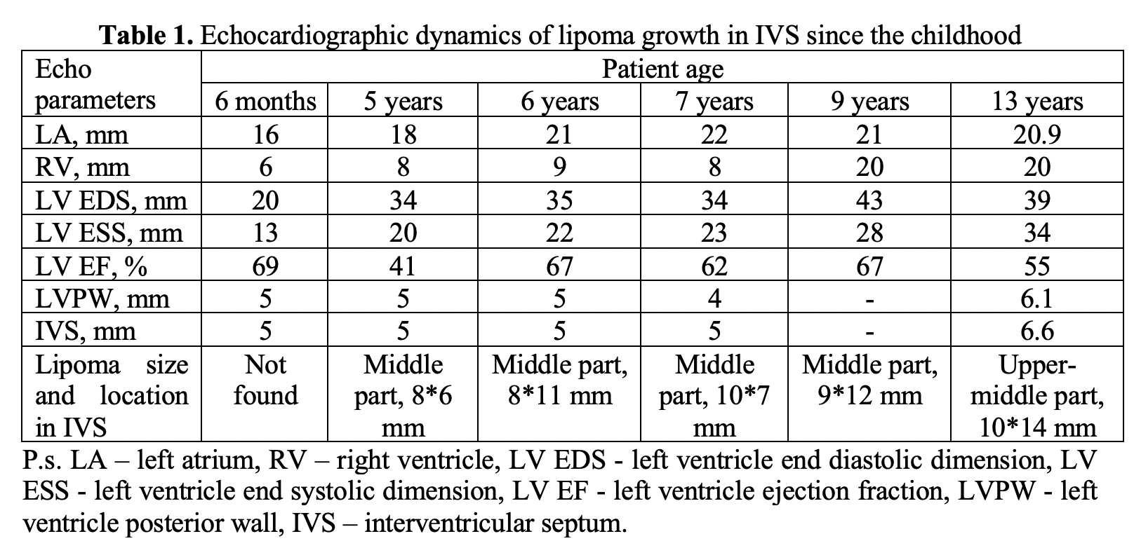
The dynamics EchoCG investigation are presented in Table 1. In 6 months, the lipoma was not found and firstly appeared in the middle part of interventricular septum (IVS) in 5 years age about 8*6 mm in size. It was growing slowly and in 13 years in the recent EchoCG the lipoma size reached 10*14 mm and was located not only in the middle, but also in upper part of interventricular septum (IVS).
Relevant Catheterization Findings
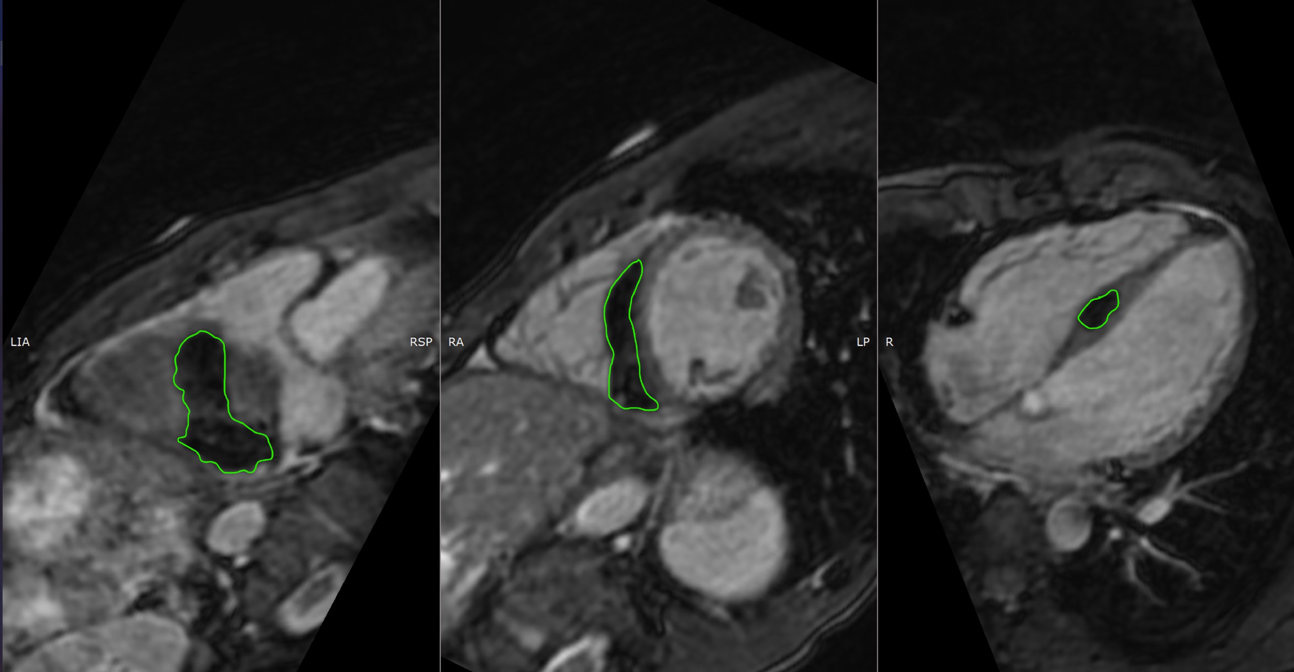
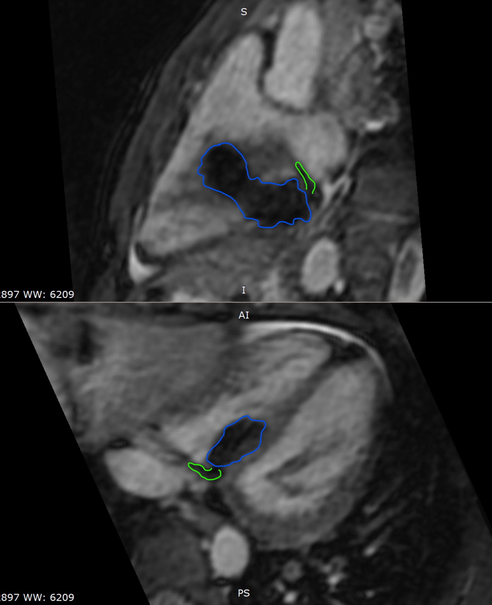
In August 2020, the MRI on GE 450w cardiac MRI in Fat Suppression Mode revealed lipoma also in the upper and middle thirds of IVS, in the RV. The structure irregular elongated shape, 49*28 mm in size and 8,77 ml in volume and consisted of adipose tissue (Fig. 1-3). In the middle of the short axis of the heart, the structure is located at the level of 9 and partially of 8 segments, in the basal parts of the short axis - at the border of 3 and 4 segments, in posterior IVS region
Interventional Management
Procedural Step
1. What is the morphology of the tumor?
While lipomas appear to not enhance and have homogenous high signal intensity on T1 and T2 weighted images, liposarcomas typically contain solid, enhancing components intermixed with areas of fat signal. Another differential diagnosis for a fat-containing mass within the heart is lipomatous hypertrophy of the interatrial septum. Lipomatous hypertrophy is a misnomer as the pathophysiology involves hyperplasia of adipocytes as opposed to hypertrophy. Unlike lipomas, these are unencapsulated, spare the fossa ovalis and thus bear a dumbbell shape.
2. Is it necessary to perform a diagnostical biopsy?
Considering the high specificity of the MRI fat suppression regimen and the location of the lipoma in the IVS, which is accompanied by the risk of intraoperative and postoperative complications for the patient, the diagnostic biopsy for now is not indicated.
3. When should a surgical resection be performed?
In the patients till the present time no pericarditis, tumor invasion, damage to adjacent structures, rapid growth, impaired cardiac hemodynamics (obstruction) and arrhythmias, this structure can be regarded as benign, are present and that is why it does not require resection. Surgical treatment will be indicated if clinical symptoms of heart failure or clinically symptomatic arrhythmia appear. It is important to note that in the case of obstruction, given the location of the lipoma in the IVS, the scope of the operation will not be radical.
Case Summary
Based on the results of the examination, we decided that there was no need for a diagnostic biopsy and consequently, surgical resection of the tumor. At the same time, recommendations were given to monitor the patient's complaints, ECG, and to conduct an MRI examination once a year to prevent dysfunction of the heart and estimate the right time for possible surgical resection in the future.