Imad Sheiban
Pederzoli Hospital, Italy
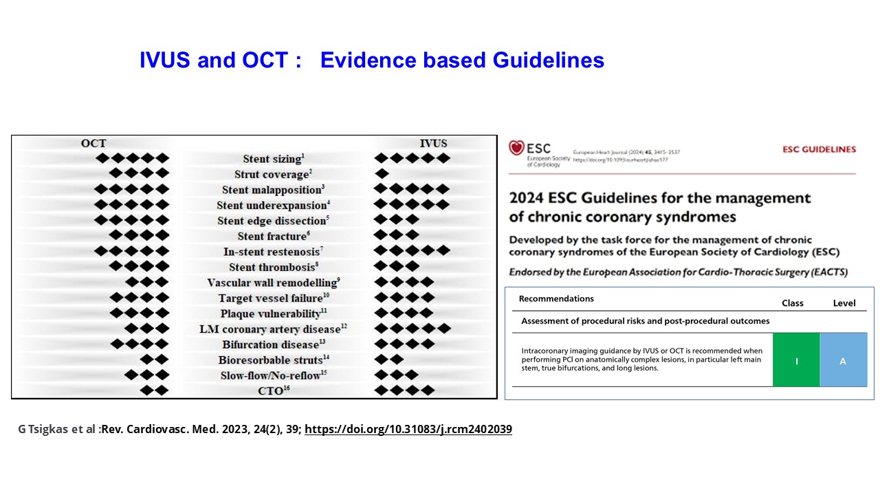
At TCTAP 2025, in the breakfast session of 'Imaging & Physiology I', Dr. Imad Sheiban (Pederzoli Hospital, Italy) addressed the evolving role of intravascular imaging in bifurcation PCI. He emphasized that angiography alone is often insufficient to guide complex bifurcation interventions. Instead, intravascular ultrasound (IVUS) and optical coherence tomography (OCT) have become essential tools to evaluate plaque composition, lesion morphology, and optimize procedural results.
IVUS and OCT provide valuable insights such as vessel sizing, plaque burden, stent expansion, malapposition, edge dissection, and side branch jailing. Longitudinal vessel imaging can also help predict carinal shift and side branch compromise. Dr. Sheiban illustrated how OCTĪ»s superior resolution detects subtle stent malapposition and edge dissections, whereas IVUS offers better vessel wall visualization in larger vessels.
Recent randomized trials and meta-analyses, including the OCTOBER and OCTIVUS studies, have confirmed that both IVUS- and OCT-guided bifurcation PCI improve clinical outcomes compared to angiography guidance alone. Both modalities are safe and effective, with no clear superiority.
In conclusion, Dr. Sheiban noted that the decision to use IVUS or OCT should be individualized based on clinical context, lesion complexity, and operator experience. Ī░The use of intravascular imaging must be incorporated into daily practice for optimal bifurcation PCI outcomes,Ī▒ he stated.
Imaging & Physiology I
Thursday, April 24, 7:00 AM-8:00 AM
Presentation Room 1, Level 1
Edited by
Hoyun Kim, MD
Bucheon Sejong Hospital, Korea (Republic of)