
Lots of interesting abstracts and cases were submitted for TCTAP 2025. Below are the accepted ones after a thorough review by our official reviewers. Don’t miss the opportunity to expand your knowledge and interact with authors as well as virtual participants by sharing your opinion in the comment section!
TCTAP C-055
Maximizing Resources or Exceeding Safety? Vessel Perforation During Extended Intravascular Lithotripsy in an Elderly STEMI Patient
By Heng Shee Kim, Wee Pang Ng, Kulasingham Vicknesan, Hou Tee Lu, Mahadevan Gurudevan
Presenter
Heng Shee Kim
Authors
Heng Shee Kim1, Wee Pang Ng1, Kulasingham Vicknesan1, Hou Tee Lu1, Mahadevan Gurudevan1
Affiliation
Sultanah Aminah Hospital, Malaysia1,
View Study Report
TCTAP C-055
Coronary - Complex PCI - Calcified Lesion
Maximizing Resources or Exceeding Safety? Vessel Perforation During Extended Intravascular Lithotripsy in an Elderly STEMI Patient
Heng Shee Kim1, Wee Pang Ng1, Kulasingham Vicknesan1, Hou Tee Lu1, Mahadevan Gurudevan1
Sultanah Aminah Hospital, Malaysia1,
Clinical Information
Patient initials or Identifier Number
Relevant Clinical History and Physical Exam
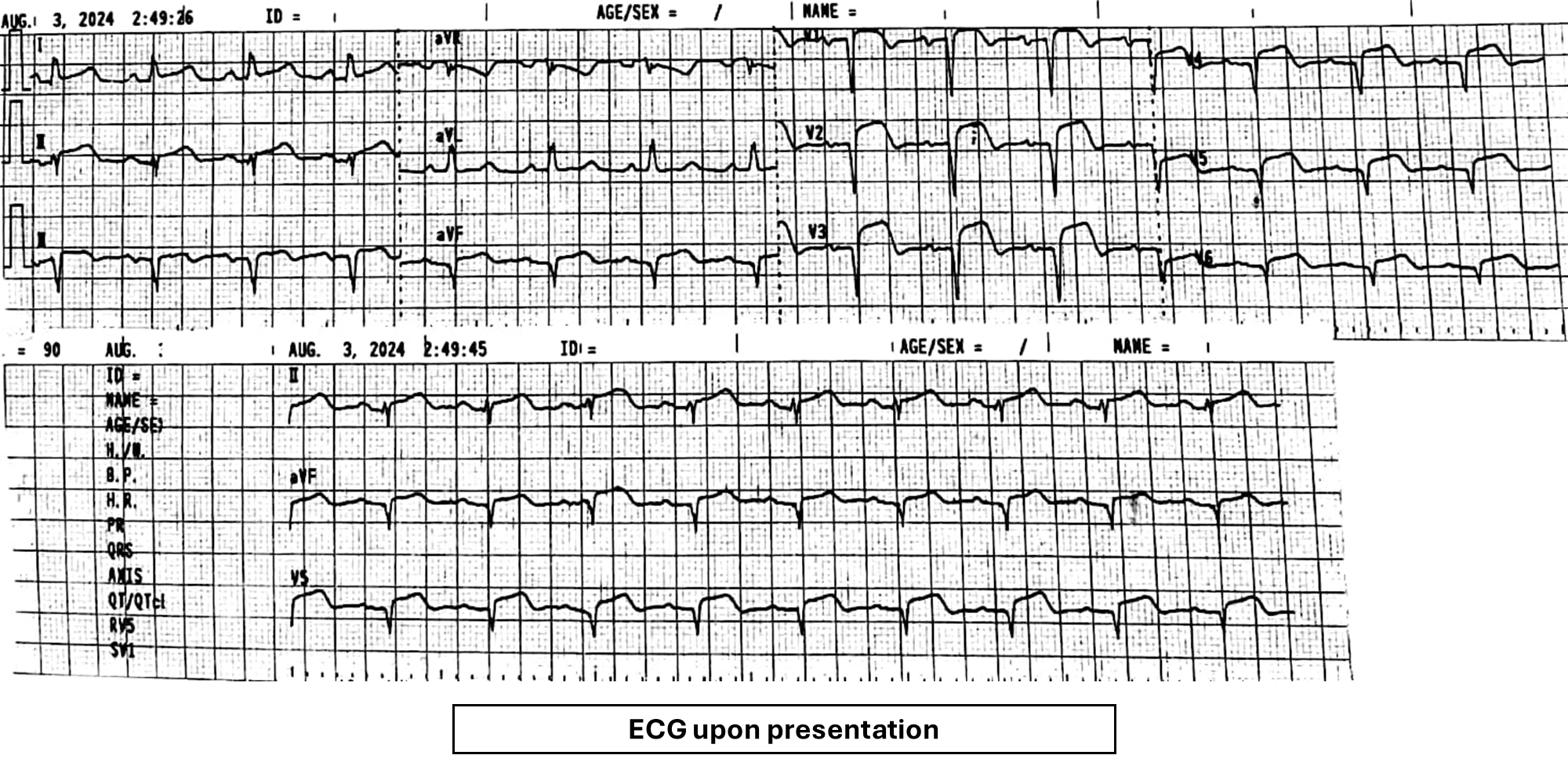
An 81-year-old functionally independent woman with Type 2 Diabetes, Hypertension, and Atrial Fibrillation presented with central chest pain and shortness of breath. Diagnosed with acute anterolateral STEMI, Killip I, she underwent fibrinolysis at a district hospital and referred to our PCI center due to persistent symptoms. Upon examination, she was hemodynamically stable with BP 125/60 mmHg, HR 89 bpm, and SpO2 100% on room air. Cardiopulmonary examination was unremarkable.
Relevant Test Results Prior to Catheterization
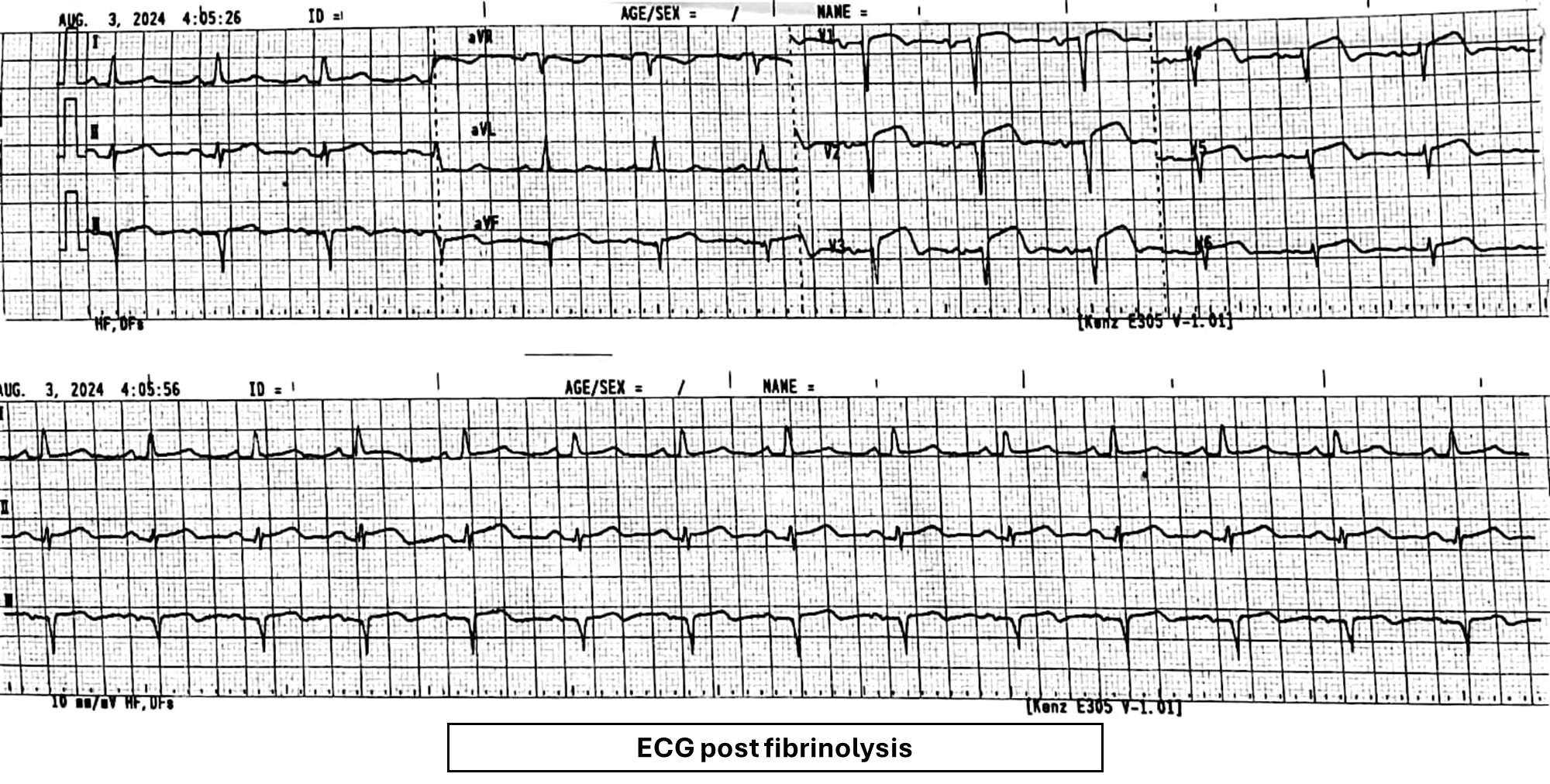
High-sensitive Troponin I during admission: 1890.6 ng/LCreatinine 156 umol/L, eGFR27 mL/min/1.73 m²LDL Cholesterol 2.8 mmol/LHbA1c: 6.4 mmol/LEchocardiogram: Poor left ventricular ejection fraction of 30 - 35%, mildly dilated left atrium and ventricle; global mild hypokinesia, functional all valve with mild mitral regurgitation.Chest x-ray: Mild cardiomegalyECG on presentation: ST elevation anterolateral leads, Q waves anterior leads; post-fibrinolysis: >50% resolution of ST-elevation.
Relevant Catheterization Findings
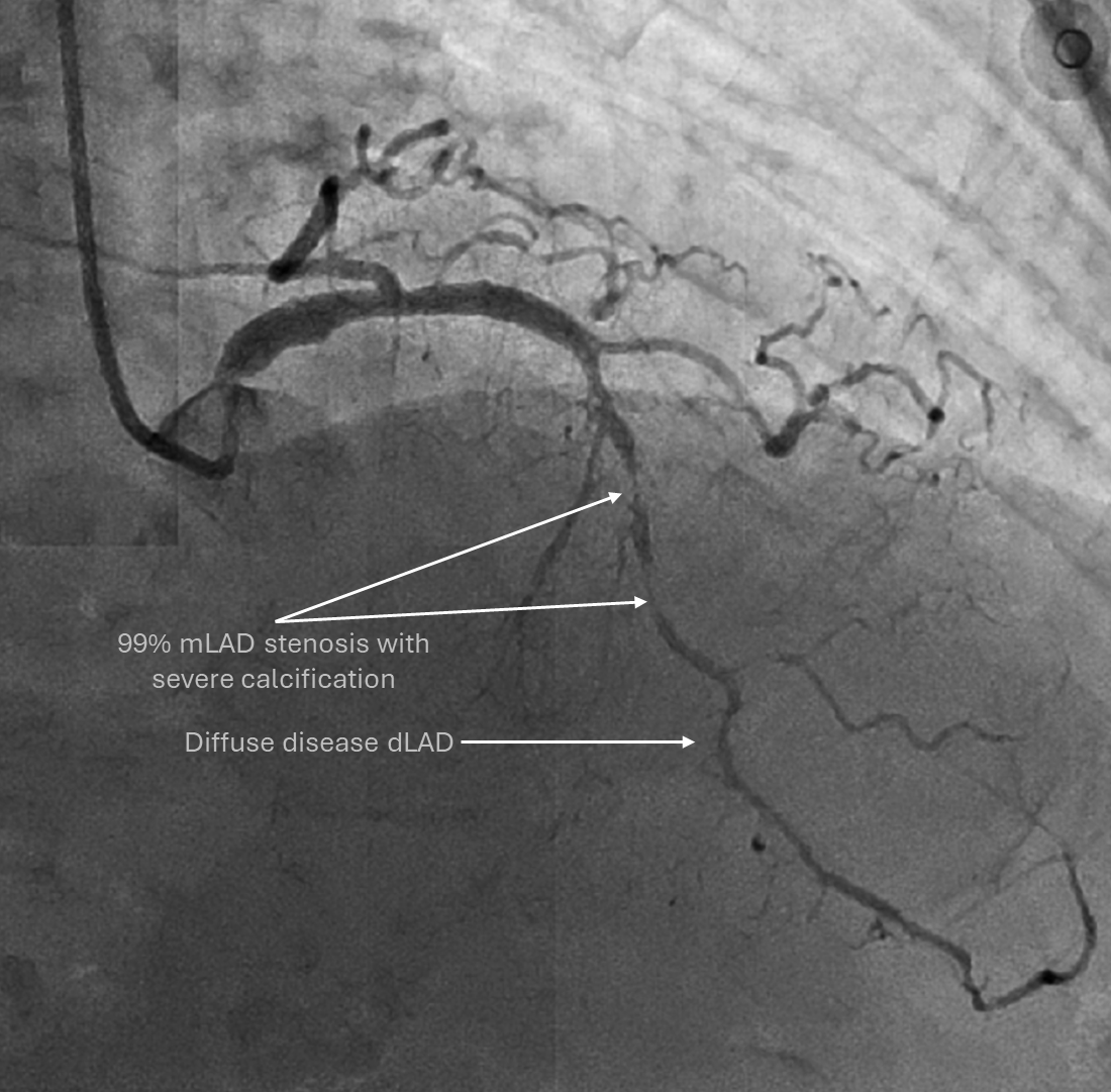
CAG revealed a normal LM, mild disease in the proximal LAD, and severe diffuse calcification, 99% stenosis in the mid-LAD, extending to the distal wrap-around LAD. The LCx was recessive with 80% stenosis in the mid LCx, and the dominant RCA showed 80% stenosis at the proximal PDA. SYNTAX score was 18, with a PCI SYNTAX II score of 63 (4-year mortality 68.4%) vs. CABG 41 (16.3%). After heart team and family discussions, PCI to LAD under IVUS guidance was chosen due to age and persistent symptoms.
Interventional Management
Procedural Step
Access & Guide: 6F Radial EBU 3.0
LAD wired with a workhorse wire. Due to the tight mLAD stenosis, lesion was prepared with 2.0 x 15 mm balloon at 6atm, followed by IVUS. IVUS showed severe 360° calcification from the mid to pLAD, with MLA of 2.88 mm² after pre-dilatation. The total calcified length was 26mm, with calcium score of 3.
She was monitored in CCU, and repeat echo showed no effusion progression. She was discharged well after two days.
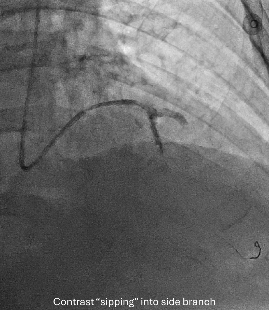
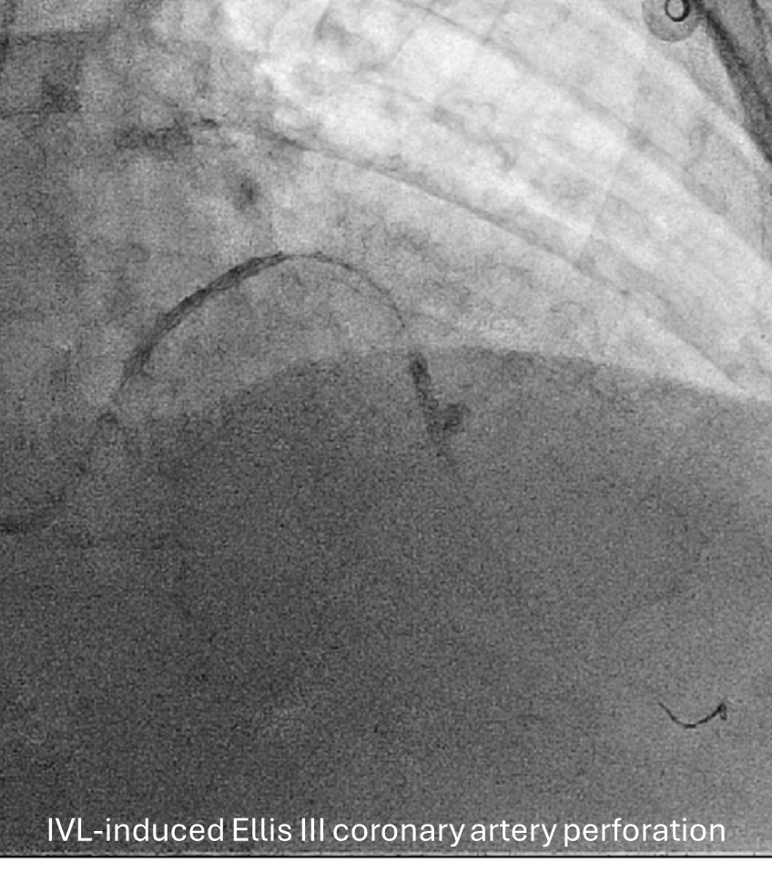
Case Summary
This case highlights the balance between resource utilization and procedural safety during IVL-guided PCI in heavily calcified lesions. While attempting to maximize the remaining IVL pulses in an elderly patient with symptomatic CAD, an Ellis Type III coronary perforation occurred. Rapid identification and deployment of a covered stent restored vessel integrity and stabilized the patient. This case emphasizes the importance of adherence to IVL protocols and prompt management of complications to achieve favorable outcomes, even in high-risk interventions.