E-Case

JACC

Lots of interesting abstracts and cases were submitted for TCTAP 2025. Below are the accepted ones after a thorough review by our official reviewers. Don¡¯t miss the opportunity to expand your knowledge and interact with authors as well as virtual participants by sharing your opinion in the comment section!

TCTAP C-125

Complex and High-Risk Intervention in Indicated Patients (Chip) - PCI for 2-Vessel CTO and Left Main Trunk Lesion With Severe Calcification in a Polyvascular Disease Patient With Cardiogenic Shock

By Yasunori Inoguchi, Ryo Nishikawa, Yohei Yakuta, Taiji Yoshida, Hidenobu Terai

Presenter

Yasunori Inoguchi

Authors

Yasunori Inoguchi1, Ryo Nishikawa1, Yohei Yakuta1, Taiji Yoshida1, Hidenobu Terai1

Affiliation

Kanazawa Cardiovascular Hospital, Japan1,
View Study Report
TCTAP C-125
Coronary - Complex PCI - Multi-Vessel Disease

Complex and High-Risk Intervention in Indicated Patients (Chip) - PCI for 2-Vessel CTO and Left Main Trunk Lesion With Severe Calcification in a Polyvascular Disease Patient With Cardiogenic Shock

Yasunori Inoguchi1, Ryo Nishikawa1, Yohei Yakuta1, Taiji Yoshida1, Hidenobu Terai1

Kanazawa Cardiovascular Hospital, Japan1,

Clinical Information

Patient initials or Identifier Number

Relevant Clinical History and Physical Exam

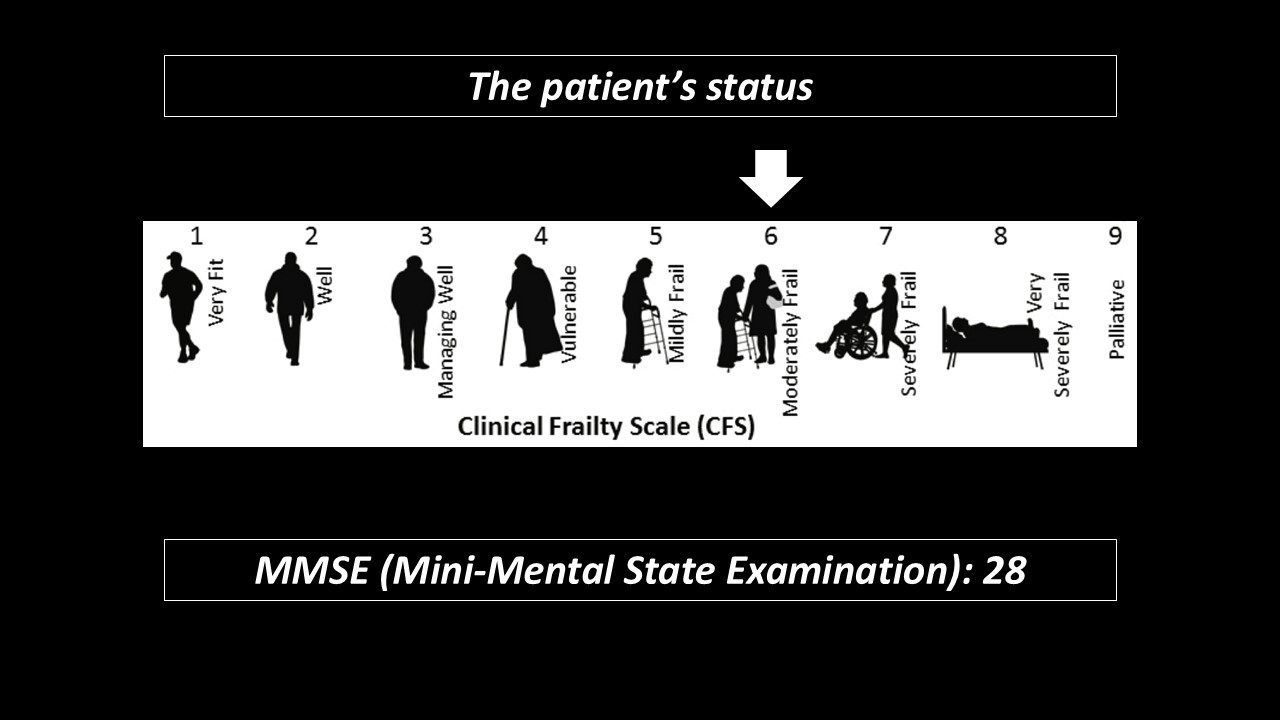
A 83-year-old woman was admitted ourhospital due to acute decompensated heart failure.She did not have obvious dementia, but herADL (Activities of Daily Living) was decreased, and her clinical frailty scalewas 6 that means moderate frailty.After admission, we treated her withoptimal medical therapy but her clinical course had not been improved.Her electrocardiogram(ECG) and echocardiography had suggested severe ischemia of myocardium.

Relevant Test Results Prior to Catheterization

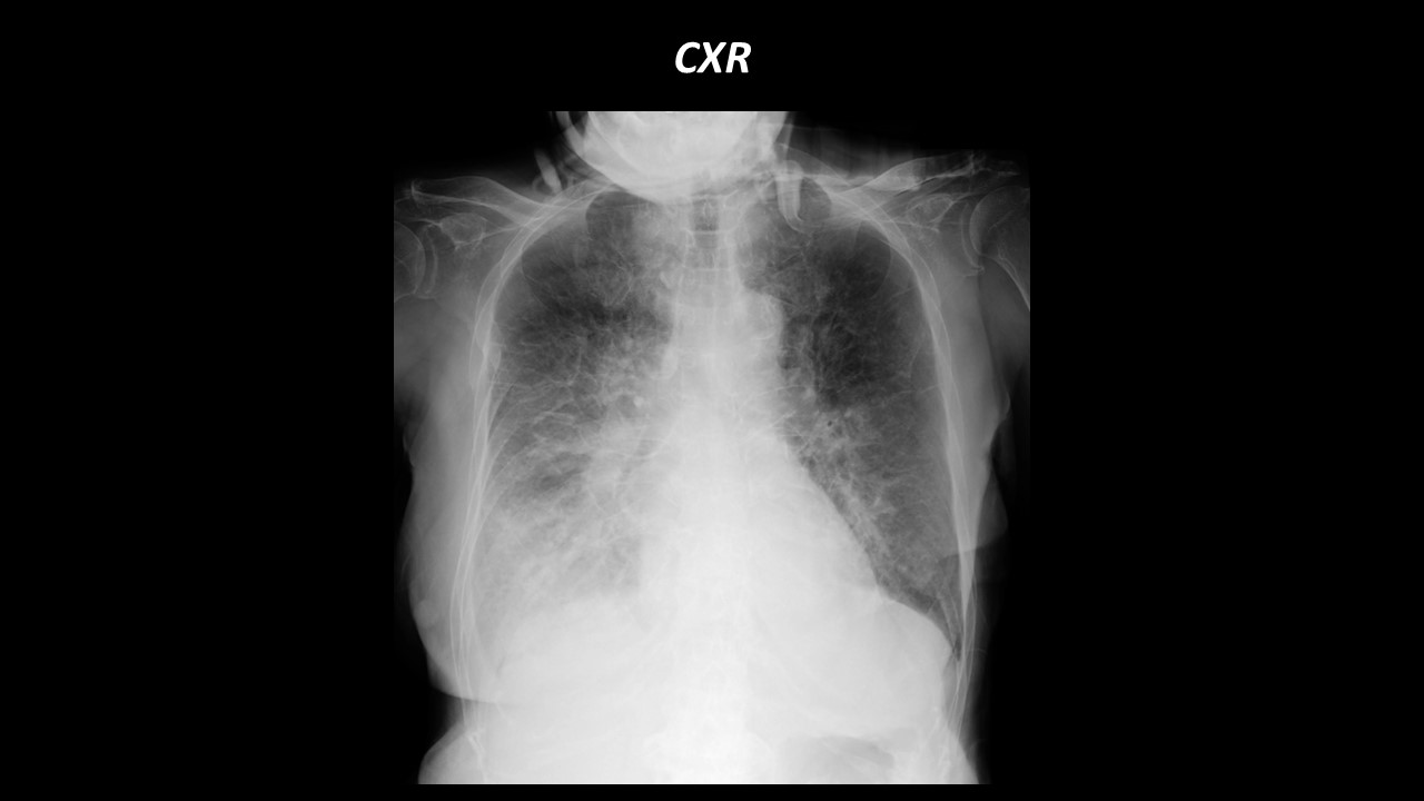
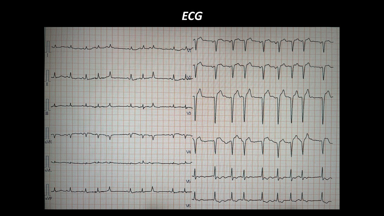
Chest X-ray showed bilateral lung congestion and pleural effusion.Electrocardiography showed poor progressionR in V1 to V4 inductions and ST depression in V5-6 induction.Echocardiography revealed ejection fractionof left ventricle (LV) was 25% with thinning of anterior wall of LV.Carotid artery ultrasound examination foundbilateral carotid artery severe stenosis.Laboratory data demonstrated elevation of serumBNP (677.9 pg/mL) and high sensitive Troponin T (0.059 ng/mL).


Relevant Catheterization Findings

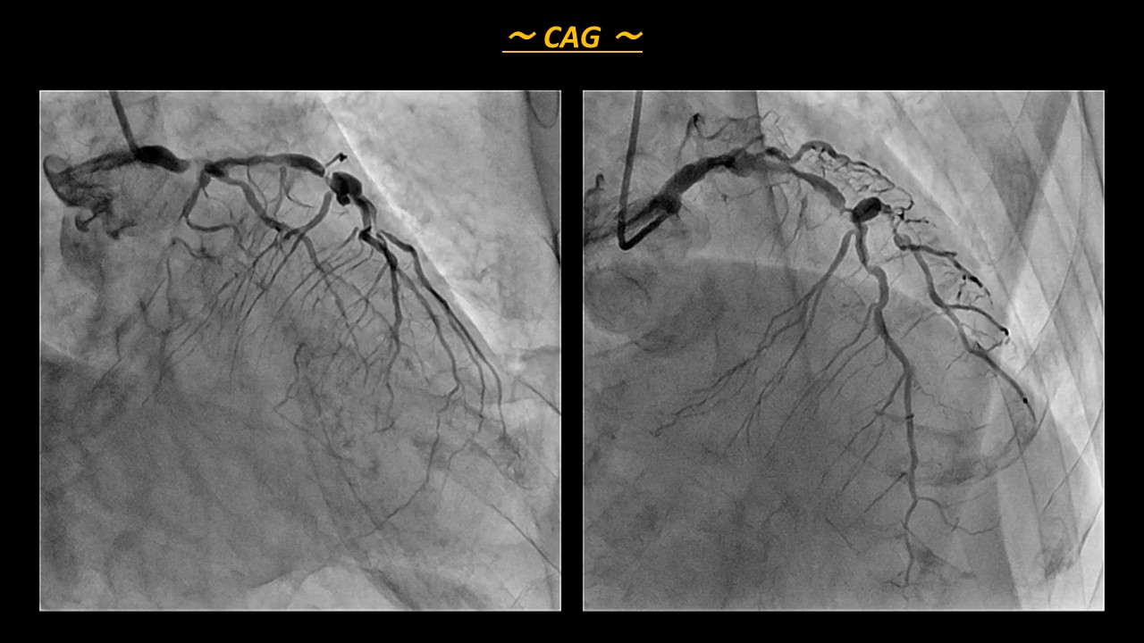
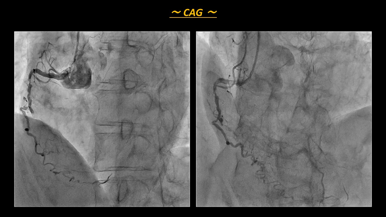
Coronary angiography (CAG) showed triplevessel disease with 2 vessel CTO. There was 99% stenosis with severecalcification in LMT, 99% stenosis in LAD seg7, and CTO in LCx seg11 and RCAseg2.After angiography, we considered how torevascularization for this patient in our heart team including cardiac surgeon.However, surgeon hesitated to CABG because of her severe comorbidities.The day after CAG, she fell into cardiogenic shock, so we had no choice but to performed emergent revascularization.

Interventional Management

Procedural Step

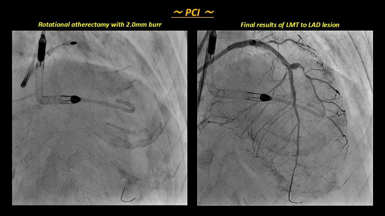
At first, we inserted the Impella CP, and triedto revascularization for LMT and LAD lesion.IVUS findings revealed concentric severecalcification in LMT lesion, because we performed rotational atherectomy byusing 1.75mm and 2.0mm burr.After that, we implanted the DES from LMTto LAD seg7, and gained enough expand the lumen.Furthermore, we negotiated RCACTO lesion and selected the strategy of retrograde approach first.Because RCA CTO was long CTO and unknownvessel route, we considered that it is absolutely necessary to establishbi-directional approach.We could success epicardial channeltracking from diagonal branch, and punctured distal cap of the CTO by Gaianext3(Asahi Intecc, Japan).However, Gaia next3 advanced outside thevessel and other wires also could not puncture distal cap of the CTO correctly becauseof severe calcified plaque in CTO.Then, we tried antegrade approach, butantegrade wiring was also difficult because of severe calcification.Antegradewire finally advanced out of vessel, furthermore, even knuckle wire technique wasnot effective.We finally broke through this nightmaresituation with scratch and go technique by antegrade approach, and make a largeshape of knuckle wire in the subintimal space at the end of CTO lesion.Bydoing that, we could retrograde puncture easily, and retrograde wire was intothe same subintimal space.We finally could establish r-CART, and successfulrevascularization of RCA CTO.


Case Summary

We finally could success the one stagerevascularization both LMT to LAD lesion and RCA CTO supported by Impella CP.This case is CHIP, and CHIP is known to becompose of three factors (patient factors, complicated heart disease, complexPCI).Among CHIP factors, unstable hemodynamics and frailty like this case wereassociated major complications.Impella CP is definitely effective in CHIPPCI but also it is reported VA-ECMO or combination with Impella and VA-ECMO inducehigh mortality and adverse event.In CHIP PCI like this case, strategy ofearly introduction of Impella and avoidance of ECMO might reduce perioperativecomplications and improve patient outcomes.