
Lots of interesting abstracts and cases were submitted for TCTAP 2025. Below are the accepted ones after a thorough review by our official reviewers. Don’t miss the opportunity to expand your knowledge and interact with authors as well as virtual participants by sharing your opinion in the comment section!
TCTAP C-195
A Challenging Case Report of Coronary Embolism Originating From an Aortomitral Intervalvular Fibrosa Pseudoaneurysm in a Patient With Infective Endocarditis, Status Post Aortic Valve Replacement and Mitral Valve Repair
By Kittichok Jonggonsuwan, Wittawat Wattanasiriporn
Presenter
Kittichok Jonggonsuwan
Authors
Kittichok Jonggonsuwan1, Wittawat Wattanasiriporn1
Affiliation
Rajavithi Hospital, Thailand1,
View Study Report
TCTAP C-195
Coronary - Imaging & Physiology - Invasive Imaging (IVUS, OCT, NIRS, VH, etc)
A Challenging Case Report of Coronary Embolism Originating From an Aortomitral Intervalvular Fibrosa Pseudoaneurysm in a Patient With Infective Endocarditis, Status Post Aortic Valve Replacement and Mitral Valve Repair
Kittichok Jonggonsuwan1, Wittawat Wattanasiriporn1
Rajavithi Hospital, Thailand1,
Clinical Information
Patient initials or Identifier Number
Relevant Clinical History and Physical Exam
A Thai 29 year-old man with history of Severe AR and severe MR due to Infective endocarditis S/P Bileaflet Aortic valve replacement with Mitral valve repair in 2022 presented to emergency department with acute chest pain 4 hour. He had history of discontinuation of warfarin 3 months prior to presentation. V/S: T 36.2 C, BP 120/66 mmHg, PR 85 bpm, RR 17 /min.Heart : Seen surgical scar at midchest, no murmur, valve’s click heard at aortic area, Lung : clear both lungs.
Relevant Test Results Prior to Catheterization
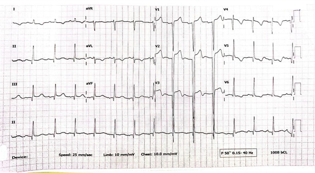
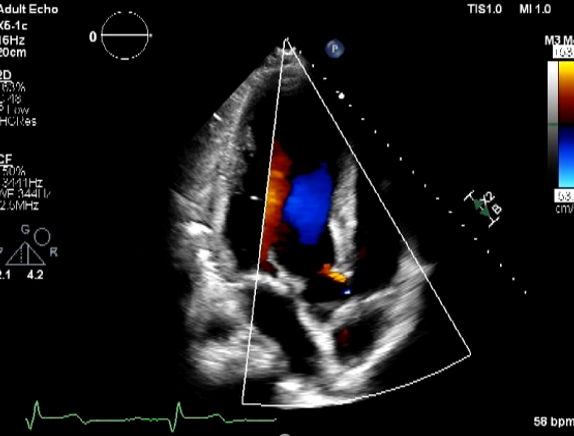
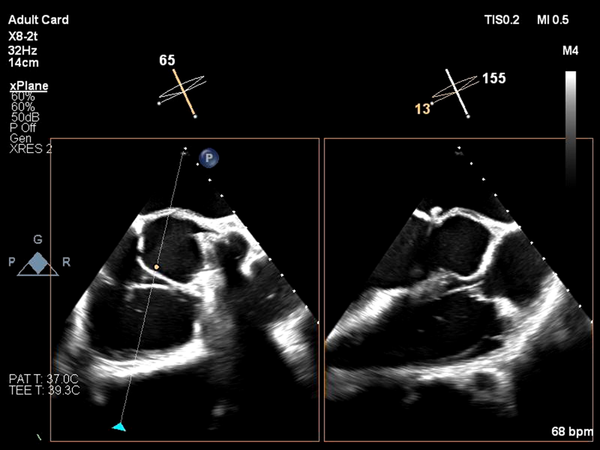
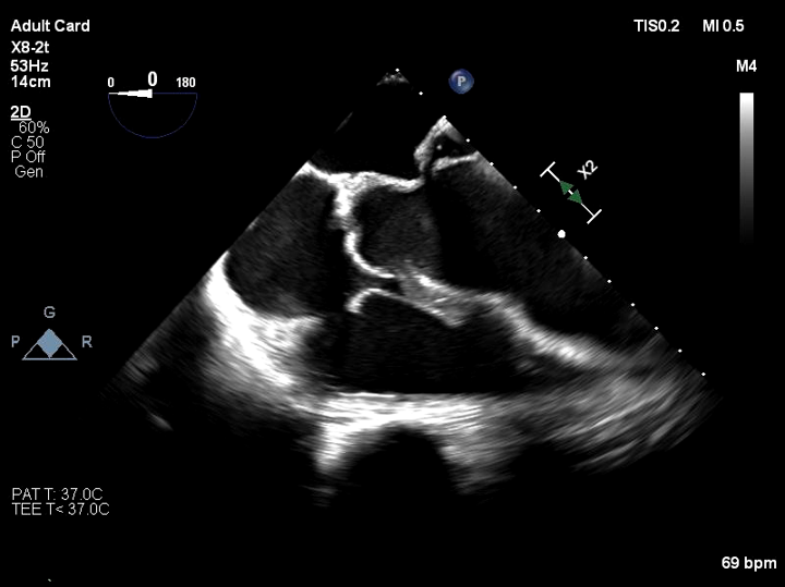
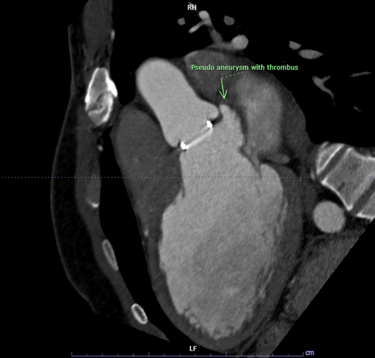
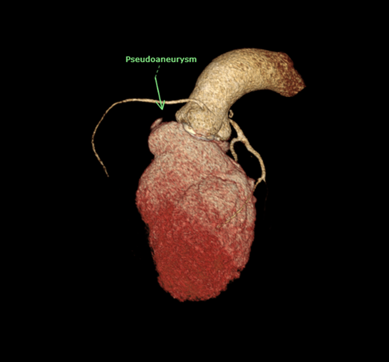
EKG showed normal sinus rhythm with ST segment elevation at V1-3 , inverted T wave at V4-6 , I ,aVL. Echocardiogram showed impaired LV systolic function, global wall hypokinesia, normal prosthetic aortic valve function with mild paravalvular leakage and bulging of aortomitral intervalvular fibrosa, suspected Aortomitral intervalvular fibrosa aneurysm. CT cardiac revealed pseudoaneurysm of the aortomitral intervalvular fibrosa with laminated thrombus with no LAA thrombus visualized.
Relevant Catheterization Findings
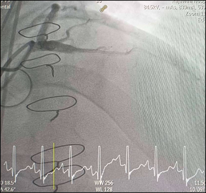
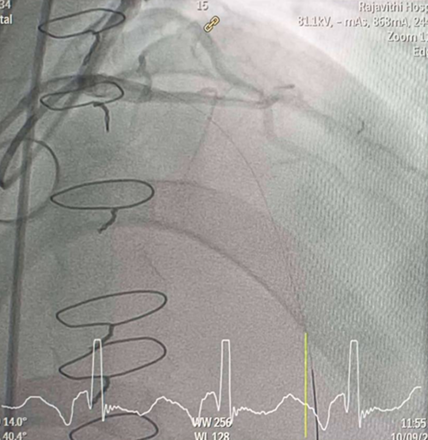
CAG was performed with right femoral approach with JL 4.0/7 Fr, JR 4.0/6 Fr diagnostic catheter showedRight dominantLM : mild irregular, non significant stenosis.LAD : total occlusion mLAD.LCx : mild irregular, non significant stenosis.RCA : non significant stenosis.
Interventional Management
Procedural Step
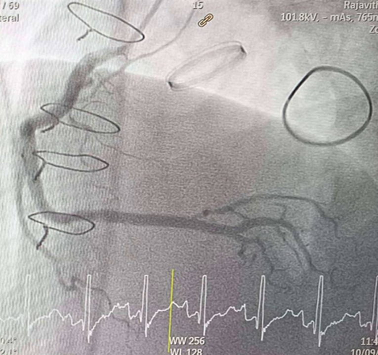
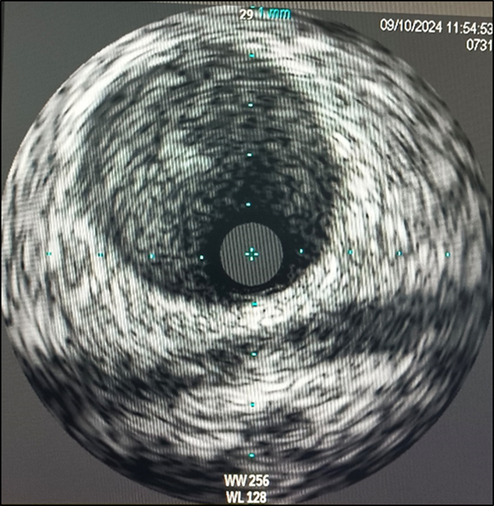
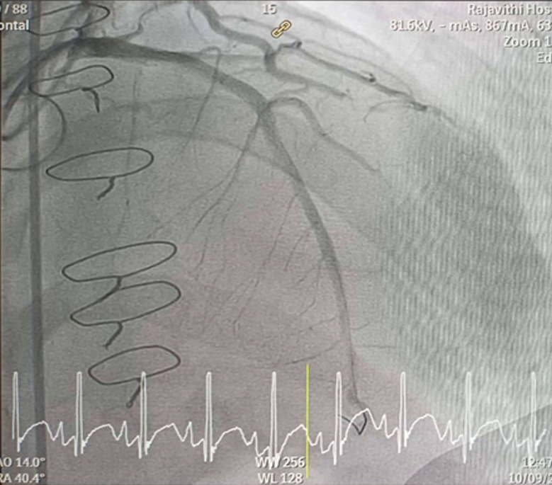
Inserted Sion wire and thrombuster to LAD revealed multiple red clots.SC balloon 2.0 x 15 mm, then 2.5 x 15 mm, was inflated at mLAD up to 12 atm. IVUS insertion to LAD revealed vessels’ size LM 5-5.5 mm, pLAD 4-4.5 mm, mLAD 3-3.5 mm with intraluminal thrombus, dLAD 2.75-3 mm. Multiple inflations were done at mLAD with SC balloon 2.5 x 15 mm. After multiple SC balloon inflations, the CAG revealed slow flow phenomenon. Then, the operator performed thrombus aspiration, multiple SC balloon 2.0 x 15 mm inflation and gave IC integrillin 3.4 ml. The final angiogram was acceptable result. The anticoagulant was given after CAG. Chest pain was subsided and hemodynamic parameters remained stable. EKG recorded after angiography showed resolution of ST elevation at V1-3. Later, the patient was discharged with warfarin. The Aortomitral intervalvular fibrosa repair surgery was planned due to embolic complication.
Case Summary
1. Coronary embolism can be considered as a rare cause of STEMI.2. Diagnosis of coronary embolism remains challenging, requires a high level of suspicion, comprehensive angiographic evaluation and other imaging modalities.3. Anticoagulant after prosthetic valve implantation should be continue lifelong to prevent further thromboembolic events.4. We report an extremely rare case of Aortomitral intervalvular fibrosa pseudoaneurysm with coronary embolism.Due to its very low incidence, it presents both a diagnostic and therapeutic challenge.5. Intravascular imaging is beneficial indeterminating mechanism of CAD, informing therapeutic strategy and confirming effective treatment of PCI.