Lots of interesting abstracts and cases were submitted for TCTAP & AP VALVES 2020 Virtual. Below are accepted ones after thoroughly reviewed by our official reviewers. Don¡¯t miss the opportunity to explore your knowledge and interact with authors as well as virtual participants by sharing your opinion!
* The E-Science Station is well-optimized for PC.
We highly recommend you use a desktop computer or laptop to browse E-posters.
CASE20200921_003
| Imaging - Invasive Imaging (IVUS, OCT, spectroscopy, etc) | |
| Repetitive "Mimic In-stent Calcification" at Mid-Right Coronary Artery in Diabetes Patients on Chronic Hemodialysis | |
| Hideyuki Aoki1, Kota Yamada1, Tetsuya Ishikawa2, Taro Takeyama2, Kahoko Mori1, Masatoshi Shimura2, Yuki Kondo2, Yukiko Mizutani1, Hidehiko Nakamura1, Itaru Hisauchi1, Shiro Nakahara1, Sayuki Kobayashi1, Isao Taguchi1 | |
| Dokkyo Medical University, Japan1, Dokkyo Medical University Saitama Medical Center, Japan2, | |
|
[Clinical Information]
- Patient initials or identifier number:
T.S.
-Relevant clinical history and physical exam:
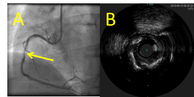
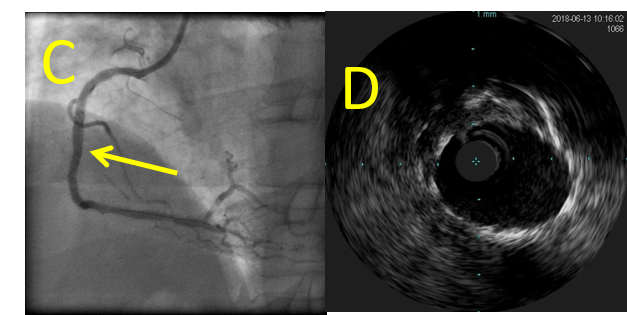
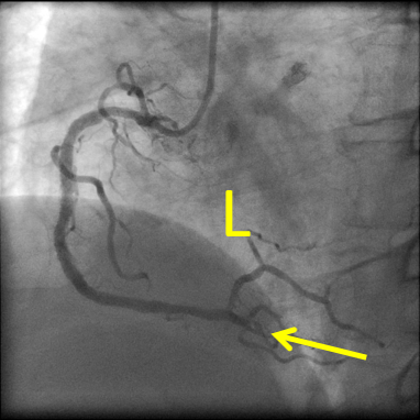
A 54-year-old male onchronic hemodialysis (HD), diagnosed as stable angina pectoris on old anteriormyocardial infarction, underwent percutaneous coronaryintervention (PCI) to diffuse moderatecalcified lesion in mid-right coronary artery (RCA) (A, B). At the initial PCI to RCA,Synergy biodegradable polymer everolims-eluting stent (3.5x38mm) was successfully placed withoutrotablator (C, D). His diabetic state was wellcontrolled, and dual antiplatelet therapy (DAPT) was continued.
  -Relevant test results prior to catheterization:
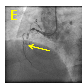
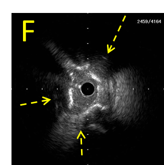
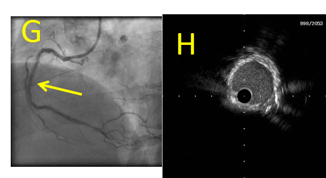
However, 8 monthlater, the secondary PCI underwent for in-stent occlusion (ISO) (E). IVUS imageat the primary stenotic site (indicated by yellow arrow in each figure) showed almostcircular superficial high-intensity layer with acoustic shadowing inside stent mimicking in-stentcalcification (ISC) (F, struts of expanded stent were indicated by yellowbroken-lined arrows). However, ISC in ISO was fully dilated by 3.5mm-sized non-compliantballoon and drug-coated balloon without indentation (G, H).
   - Relevant catheterization findings:
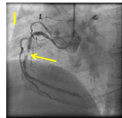
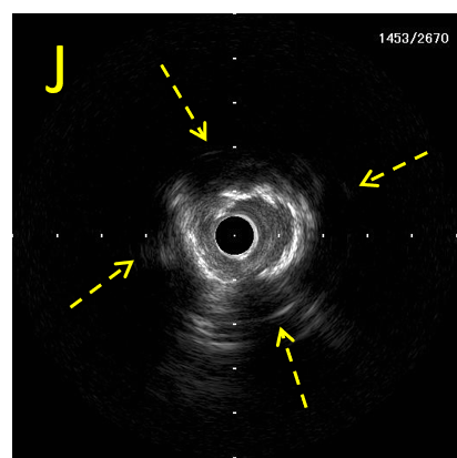
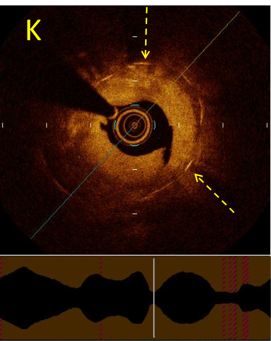
One-year later,however, severe in-stent restenosis (ISR) at the primary stenotic site was recurred(I) with recurrent superficial high-intensity layer (J). Opticalcoherence tomography (OCT) image showed layered neo-intimal pattern as high-backscattering growing mass with signalattenuation (seen in 4 to 9 o¡¯clock) representing thick neo-intima growth inside Synergy stent, without characteristicsharp bordered low-intensity presentation of calcification (K).
   |
|
|
[Interventional Management]
- Procedural step:
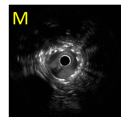
Because of the recurrence of superficialhigh-intensity layer, a biodegradable polymersirolimus-eluting stent (Orsiro 3.5 x 24 mm long) was placed inside Synergywith good expansion (L, M). ISC is usually necessary to modify by a rotablatorprior to ballooning and stenting because of severe advanced atherosclerosisdeveloped during a long-term1. The present repetitive superficial high-intensity layer complicated in early (< 12 months) ISR cases, and inpatients on HD, mainly comprised hyperplastic neo-intima easily to dilate.Therefore, we proposed the present superficial circular high intensity imagesby IVUS as ¡°mimic ISC¡± by discriminating from ISC in view of balloonexpandability (H, M) and with layered in-stent restenotic OCT image withoutcalcification (K).
  - Case Summary:
ISC isusually necessary to modify by a rotablator prior to ballooning and stenting becauseof severe advanced atherosclerosis developed during a long-term1.The present repetitive superficialhigh-intensity layercomplicated in early (< 12 months) ISR cases, and in patients on HD, mainlycomprised hyperplastic neo-intima easily to dilate. Therefore, we proposed thepresent superficial circular high intensity images by IVUS as ¡°mimic ISC¡± bydiscriminating from ISC in view of balloon expandability (H, M) and with layeredin-stent restenotic OCT image without calcification (K).
Conclusion: Since patients on HDwere the significant predictor of late (30-365 days) stent thrombosis in thepresent drug-eluting stent era, the features and the underlying mechanisms to develop¡°mimic ISC¡± during a relatively early phase should be clarified. |
|