Lots of interesting abstracts and cases were submitted for TCTAP & AP VALVES 2020 Virtual. Below are accepted ones after thoroughly reviewed by our official reviewers. Don¡¯t miss the opportunity to explore your knowledge and interact with authors as well as virtual participants by sharing your opinion!
* The E-Science Station is well-optimized for PC.
We highly recommend you use a desktop computer or laptop to browse E-posters.
CASE20200917_001
| Complications - Complications | |
| Coronary Artery Dissection After Contrast Injection Through Microcatheter | |
| Minsuk Kim1 | |
| PMC hospital, Korea (Republic of)1, | |
|
[Clinical Information]
- Patient initials or identifier number:
WCM
-Relevant clinical history and physical exam:
This 63 years old male had underwent CAG at 27th, March, 2020 as combine CAG with TFCA due to CVA.At that time, 3VD was noted, but PCI to LCX was failed. We planned follow up CAG for this patient at 11th, September, 2020.He showed no chest pain or dyspnea and his functional capacity was good.
-Relevant test results prior to catheterization:
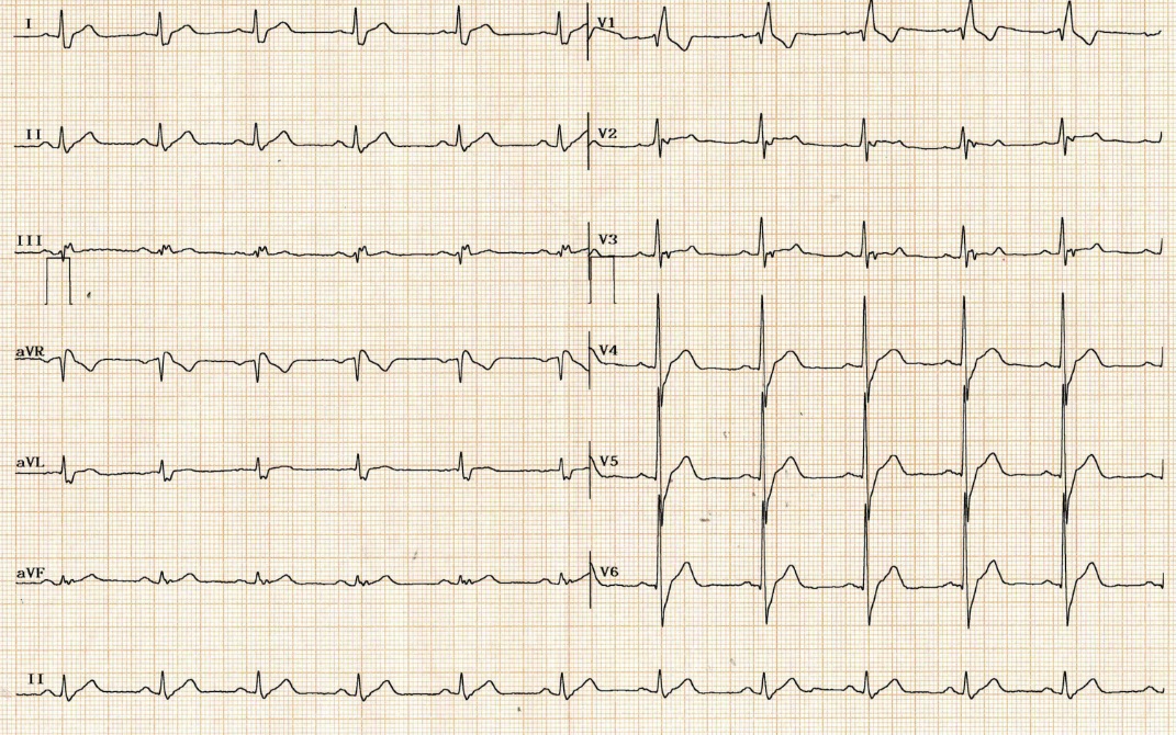
He had medications for hypertension, diabetes mellitus and dyslipidemia.His ECG showed sinus rhythm with RBBB and echocardiographic finding was tolerable with normal LVEF and no RWMA.
 - Relevant catheterization findings:
Previous CAG at March, 2020 revealed 3VD.I planned to performed PCI to OM bifurcation and diagonal branch. (video 1)I tried wiring to OM but microcatheter was not able to enter OM. (video 2)I pushed some contrast through microcatheter to see the catheter was in true lumen, but dye staining was observed and OM flow was totally occluded.I tried re-wiring to OM many times, but eventually failed wiring and decided to perform re-look CAG after 6 months. (video 3)
|
|
|
[Interventional Management]
- Procedural step:
Luckily, follow up CAG showed restored OM flow. (video 4)I tried wiring to OM from a different view than previous PCI.I used Fielder XT-R and carefully manipulated to slide in OM.As the OM os lesion had dissection before, I decided to perform culottes technique to place side branch stent first. (video 5)Two stents (Xience Sierra, 2.5 x 23mm, #2) were inserted according to conventional culottes technique protocol.Distal LCX lesion was revealed after stenting, but I decided to do medical treatment and planned follow up CAG after 6 months. (video 6)
- Case Summary:
We should not to push contrast through microcatheter to check whether the microcatheter is in the true lumen.
It can worsen coronary artery dissection, and make procedure much harder or even impossible than before. |
|