Lots of interesting abstracts and cases were submitted for TCTAP & AP VALVES 2020 Virtual. Below are accepted ones after thoroughly reviewed by our official reviewers. Don¡¯t miss the opportunity to explore your knowledge and interact with authors as well as virtual participants by sharing your opinion!
* The E-Science Station is well-optimized for PC.
We highly recommend you use a desktop computer or laptop to browse E-posters.
CASE20190915_001
| IMAGING AND PHYSIOLOGIC LESION ASSESSMENT - Imaging: Intravascular | |
| Imaging Guided CTO Intervention | |
| Yongbai Luo1, Ning Guo1 | |
| The First Affiliated Hospital of Xi'an Jiaotong University, China1, | |
|
[Clinical Information]
- Patient initials or identifier number:
1561308 LQQ
-Relevant clinical history and physical exam:
62 years female was presented with exertional chest discomfort for 1 year. 9 months ago, she was received angiography and showed that mRCA was moderate stenosis, mLAD was total occlusion and CX was severer stenosis. PCI for LAD was failed and one stent was implanted in CX. The patient was hopitalized for LAD intervention. The PE was negative.
-Relevant test results prior to catheterization:
- Relevant catheterization findings:
mLAD was total occlusion without stump, and RCA was moderate disease. Some lateral branches from RCA to LAD, but very torturous.
|
|
|
[Interventional Management]
- Procedural step:
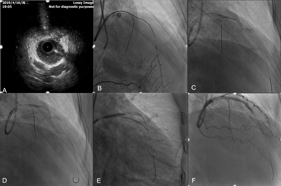
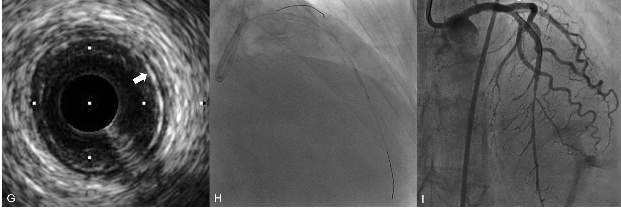
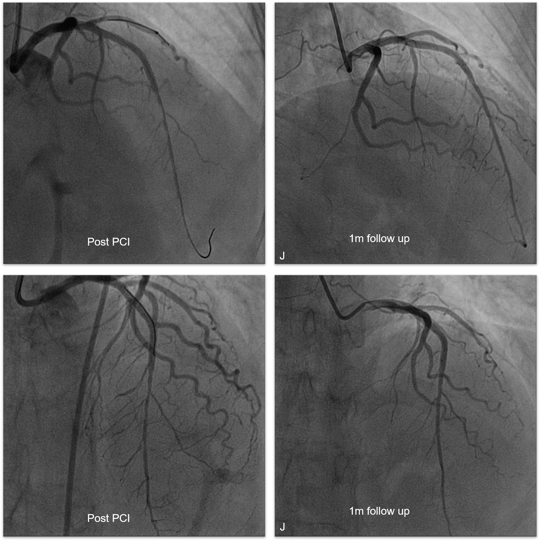
IVUS guided antegrade wiring was performed, LAD was successfully penetrated with Gaia 2nd(fig A and fig B). After predialated with 2.0 balloon(fig C), IVUS was checked to confirm Landing zone. However , after 2 stents was deployed(fig D and fig E), the angiography showed that distal segment was diffuse luminal narrowing(fig F), and IVUS was checked again and Peri-media high echo band£¨PHB£©phenomenon was confirmed in the distal reference segment£¨fig G£©, so we dilated distal LAD with 2.0 balloon with 4atm£¨fig H£©£¬and final result was acceptable£¨fig I£©£¬another unnecessary stent in the distal segmentwas avoided. And 1 month follow up angiography showed the distal LAD was enlarged£¨fig J£©.
   - Case Summary:
IVUS Guiding can simplify antegrade CTO procedure, shorten operating time, reduce the dosage of contrast medium.PHB (Peri-medium High-echoic Band) phenomenon is a predictor for late lumen gain in CTO PCI.
|
|