Lots of interesting abstracts and cases were submitted for TCTAP & AP VALVES 2020 Virtual. Below are accepted ones after thoroughly reviewed by our official reviewers. Don’t miss the opportunity to explore your knowledge and interact with authors as well as virtual participants by sharing your opinion!
* The E-Science Station is well-optimized for PC.
We highly recommend you use a desktop computer or laptop to browse E-posters.
CASE20190813_001
| CORONARY - Bifurcation/Left Main Diseases and Intervention | |
| The Result of Follow-Up After 1 Year - Left Main Bifurcation Lesion Treated by Simple Crossover Technique and IVUS Guided the LAD PCI | |
| Lyu Qiang1 | |
| Xi’an Gaoxin Hospital, China1, | |
|
[Clinical Information]
- Patient initials or identifier number:
201531589
-Relevant clinical history and physical exam:
An 80-year old man was admitted for scheduled follow-upcoronary angiography after 1 year left main bifurcation lesion treated bysimple crossover technique. Physical examination was normal. One year before, we deployed anEVERLINK stent 3.0* 28mm (12 atm, 3.15 mm) at proximal to mid LAD. Then weimplanted an EVERLINK stent 3.5 * 23 mm (14 atm, 3.84 mm) at the ostium of LMto proximal of LAD which was a simple crossover technique for LM bifurcationlesion. Two stents was overlapped.
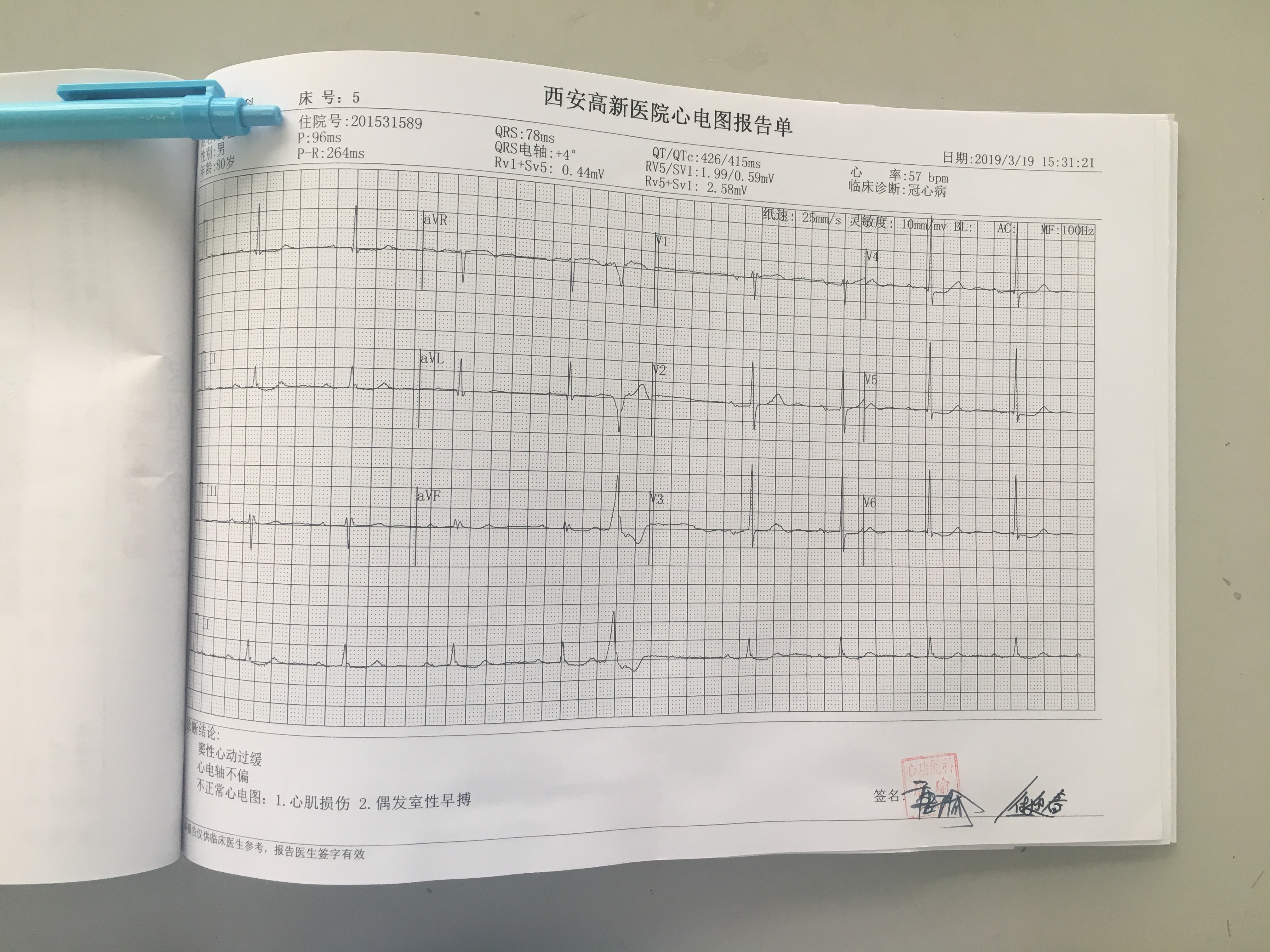
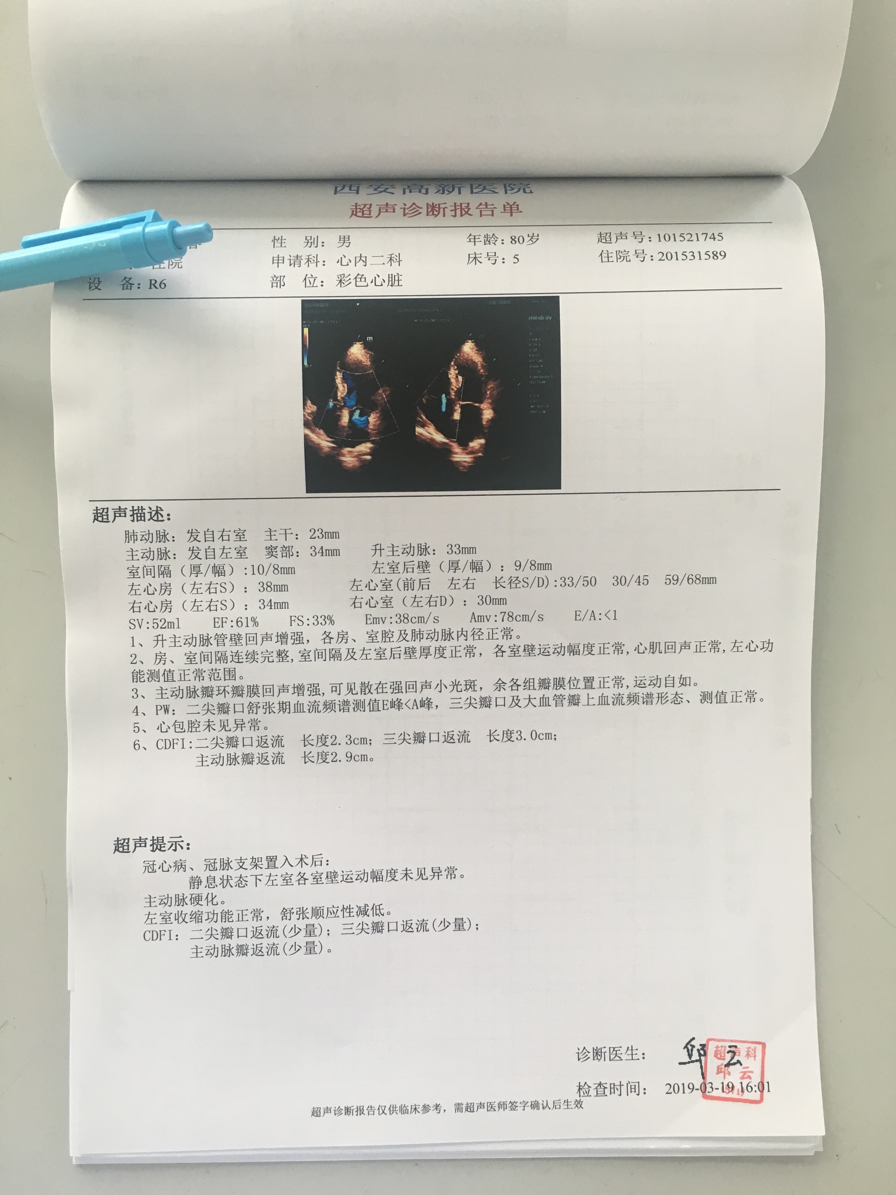
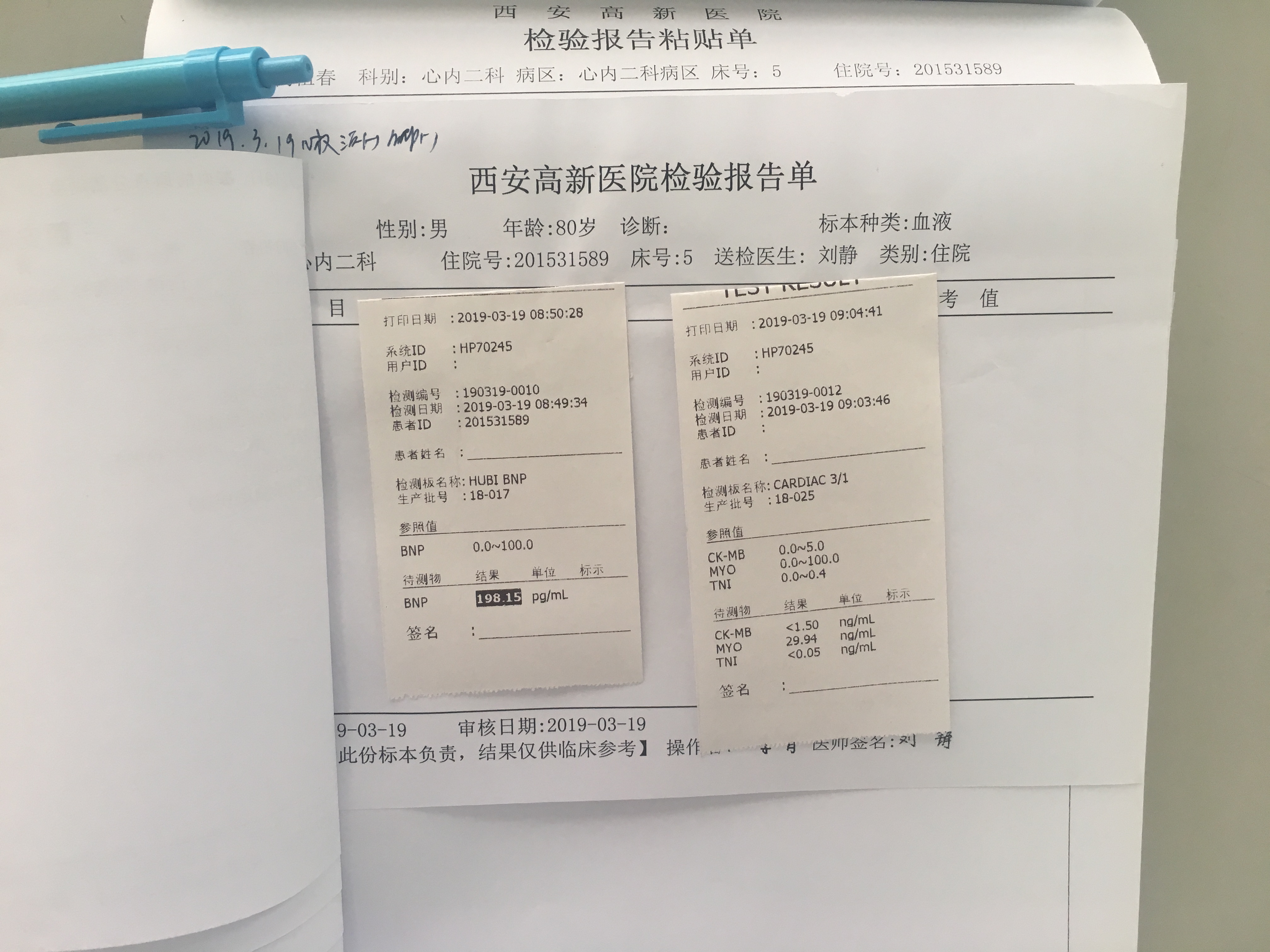
   -Relevant test results prior to catheterization:
The cardiac enzyme was unremarkable. But the BNP was 198.15 pg/ml. Hiscardiovascular risk factor was hypertension. Baseline electrocardiography(ECG)showed ST-segment reduction and T-wave inversion in V3-6, and first degree A-Vblock. Echocardiography revealed no regional wall motion abnormality and normalleft ventricular systolic fraction (EF=70%).
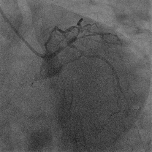
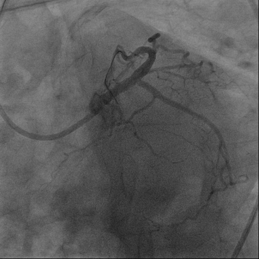
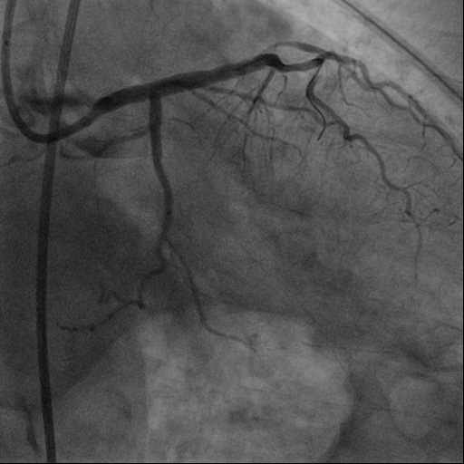
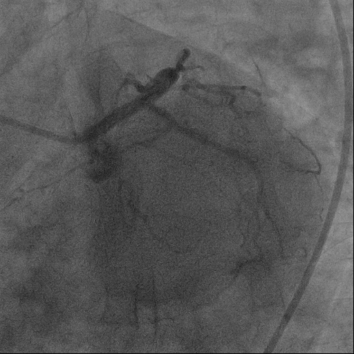
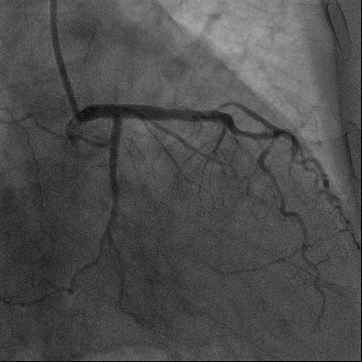
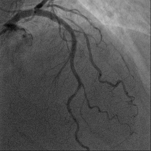
   - Relevant catheterization findings:
After 1 year, follow-up angiography showed the left coronaryangiogram was normal. The stent deployed in the proximal of LCX was patency withoutout in-stent restenosis and the blood flow was TIMI 3. The stents deployed in theLM and proximal of LAD was patency without out in-stent restenosis and the blood flowwas TIMI 3 either.
   |
|
|
[Interventional Management]
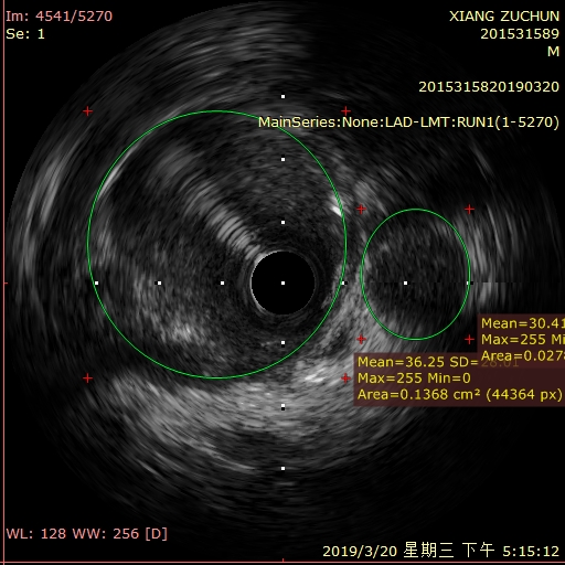
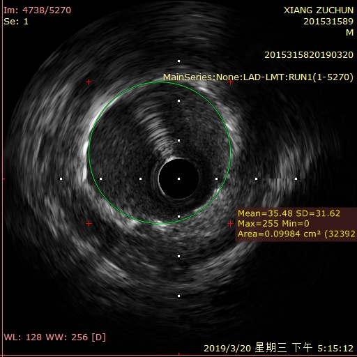
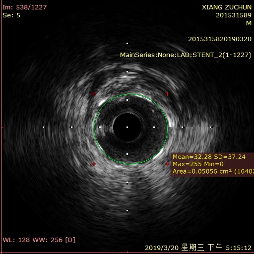
- Procedural step:
we check the IVUS.The IVUS images showed that the minimal stent area at the LM was 9.98 mm2 and the ostium of LAD was 10.99 mm2, and ostium of LCX luminalarea was 2.7mm2without significant atherosclerotic plaque at the LM bifurcation segment andthere was nosignificant neointimal hyperplasia with continuity of stent struts..The IVUS images also showed the minimal lumenal area was 2.2 mm2 at themid-LAD lesion. Then we deployed a EVERLINK 2.5×18 mm stent at the mid-LAD lesion. The finalangiogram and IVUS showed the procedure was successful.
   - Case Summary:
According theclinical symptoms of follow up 1 year, there was no MACE taking place. Theangiography and IVUS both demonstrated that the clinical outcome of the LM bifurcation lesion treated by simple crossover technique was very successful for this patient.
|
|